Refine search
Actions for selected content:
106116 results in Materials Science
Processing metallic materials far from equilibrium
-
- Journal:
- MRS Bulletin / Volume 45 / Issue 11 / November 2020
- Published online by Cambridge University Press:
- 10 November 2020, pp. 906-909
- Print publication:
- November 2020
-
- Article
-
- You have access
- HTML
- Export citation

In situ mapping of chemical segregation using synchrotron x-ray imaging
-
- Journal:
- MRS Bulletin / Volume 45 / Issue 11 / November 2020
- Published online by Cambridge University Press:
- 10 November 2020, pp. 934-942
- Print publication:
- November 2020
-
- Article
- Export citation
MRS volume 45 issue 11 Cover and Front matter
-
- Journal:
- MRS Bulletin / Volume 45 / Issue 11 / November 2020
- Published online by Cambridge University Press:
- 10 November 2020, pp. f1-f6
- Print publication:
- November 2020
-
- Article
-
- You have access
- Export citation
Solute trapping in rapid solidification
-
- Journal:
- MRS Bulletin / Volume 45 / Issue 11 / November 2020
- Published online by Cambridge University Press:
- 10 November 2020, pp. 910-915
- Print publication:
- November 2020
-
- Article
- Export citation
Glassy lithium forms superior metal anode for rechargeable Li batteries
-
- Journal:
- MRS Bulletin / Volume 45 / Issue 11 / November 2020
- Published online by Cambridge University Press:
- 10 November 2020, pp. 882-883
- Print publication:
- November 2020
-
- Article
-
- You have access
- HTML
- Export citation
2020 Virtual MRS Spring/Fall Meeting & Exhibit first of its kind for Society
-
- Journal:
- MRS Bulletin / Volume 45 / Issue 11 / November 2020
- Published online by Cambridge University Press:
- 10 November 2020, pp. 965-967
- Print publication:
- November 2020
-
- Article
-
- You have access
- HTML
- Export citation
Pioneering a new publishing model
-
- Journal:
- MRS Bulletin / Volume 45 / Issue 11 / November 2020
- Published online by Cambridge University Press:
- 10 November 2020, pp. 877-878
- Print publication:
- November 2020
-
- Article
-
- You have access
- HTML
- Export citation
MRS Journal Highlights
-
- Journal:
- MRS Bulletin / Volume 45 / Issue 11 / November 2020
- Published online by Cambridge University Press:
- 10 November 2020, pp. 880-881
- Print publication:
- November 2020
-
- Article
-
- You have access
- HTML
- Export citation
CAREER CENTRAL
-
- Journal:
- MRS Bulletin / Volume 45 / Issue 11 / November 2020
- Published online by Cambridge University Press:
- 10 November 2020, p. 975
- Print publication:
- November 2020
-
- Article
-
- You have access
- Export citation
Variation and variability
-
- Journal:
- MRS Bulletin / Volume 45 / Issue 11 / November 2020
- Published online by Cambridge University Press:
- 10 November 2020, p. 976
- Print publication:
- November 2020
-
- Article
-
- You have access
- HTML
- Export citation
EU announces European Green Deal €1 billion investment
-
- Journal:
- MRS Bulletin / Volume 45 / Issue 11 / November 2020
- Published online by Cambridge University Press:
- 10 November 2020, p. 888
- Print publication:
- November 2020
-
- Article
-
- You have access
- HTML
- Export citation
Bacterial inactivation characteristics of magnesium–calcium–zinc alloys for bone implants
-
- Journal:
- MRS Communications / Volume 10 / Issue 4 / December 2020
- Published online by Cambridge University Press:
- 30 October 2020, pp. 609-612
- Print publication:
- December 2020
-
- Article
- Export citation
Nanostructure and compositional segregation of soft magnetic FeNi-based nanocomposites with multiple nanocrystalline phases
-
- Journal:
- Journal of Materials Research , First View
- Published online by Cambridge University Press:
- 29 October 2020, pp. 1-9
-
- Article
-
- You have access
- Open access
- HTML
- Export citation

Study of the synthetic process of CaBi2Ta2O9 powder by the molten-salt method
-
- Journal:
- Journal of Materials Research , First View
- Published online by Cambridge University Press:
- 29 October 2020, pp. 1-9
-
- Article
- Export citation
Formation of micro-mechanical interlocking sites by nanoscale sculpturing for composites or hybrid materials with stainless steel
-
- Journal:
- Journal of Materials Research / Volume 35 / Issue 23-24 / 14 December 2020
- Published online by Cambridge University Press:
- 29 October 2020, pp. 3145-3156
- Print publication:
- 14 December 2020
-
- Article
- Export citation
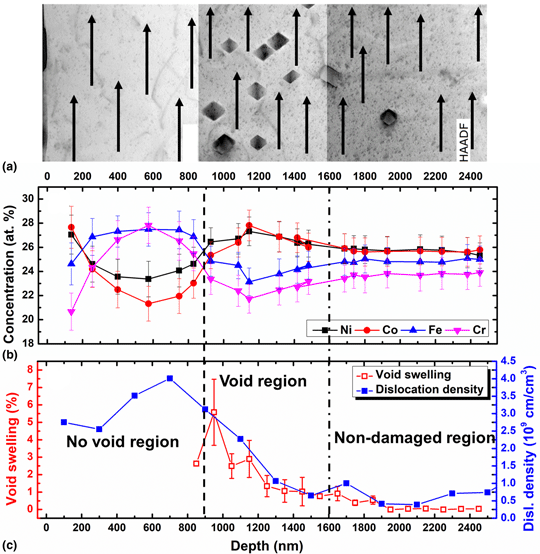
Diffusion-mediated chemical concentration variation and void evolution in ion-irradiated NiCoFeCr high-entropy alloy
-
- Journal:
- Journal of Materials Research , First View
- Published online by Cambridge University Press:
- 29 October 2020, pp. 1-13
-
- Article
-
- You have access
- Open access
- HTML
- Export citation
Double transition-metal MXenes: Atomistic design of two-dimensional carbides and nitrides - ADDENDUM
-
- Journal:
- MRS Bulletin , First View
- Published online by Cambridge University Press:
- 29 October 2020, p. 1
-
- Article
-
- You have access
- Export citation
JMR volume 35 issue 20 Cover and Back matter
-
- Journal:
- Journal of Materials Research / Volume 35 / Issue 20 / 28 October 2020
- Published online by Cambridge University Press:
- 28 October 2020, pp. b1-b2
- Print publication:
- 28 October 2020
-
- Article
-
- You have access
- Export citation
JMR volume 35 issue 20 Cover and Front matter
-
- Journal:
- Journal of Materials Research / Volume 35 / Issue 20 / 28 October 2020
- Published online by Cambridge University Press:
- 28 October 2020, pp. f1-f5
- Print publication:
- 28 October 2020
-
- Article
-
- You have access
- Export citation
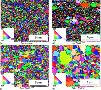
Development of advanced Fe–Al–O ODS alloy microstructure and properties due to heat treatment
-
- Journal:
- Journal of Materials Research / Volume 35 / Issue 20 / 28 October 2020
- Published online by Cambridge University Press:
- 28 October 2020, pp. 2789-2797
- Print publication:
- 28 October 2020
-
- Article
- Export citation
