Refine search
Actions for selected content:
106116 results in Materials Science

Graphene oxide coated popcorn-like Ag nanoparticles for reliable sensitive surface-enhanced Raman scattering detection of drug residues
-
- Journal:
- Journal of Materials Research / Volume 34 / Issue 17 / 16 September 2019
- Published online by Cambridge University Press:
- 12 March 2019, pp. 2935-2943
- Print publication:
- 16 September 2019
-
- Article
- Export citation
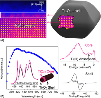
Recent progress in characterization of the core–shell structure of black titania
-
- Journal:
- Journal of Materials Research / Volume 34 / Issue 7 / 15 April 2019
- Published online by Cambridge University Press:
- 12 March 2019, pp. 1138-1153
- Print publication:
- 15 April 2019
-
- Article
- Export citation
In vitro corrosion and hemocompatibility evaluation of electrical discharge treated cobalt–chromium implant
-
- Journal:
- Journal of Materials Research / Volume 34 / Issue 8 / 29 April 2019
- Published online by Cambridge University Press:
- 12 March 2019, pp. 1363-1370
- Print publication:
- 29 April 2019
-
- Article
- Export citation
Three-dimensional biomimetic scaffolds for hepatic differentiation of size-controlled embryoid bodies
-
- Journal:
- Journal of Materials Research / Volume 34 / Issue 8 / 29 April 2019
- Published online by Cambridge University Press:
- 12 March 2019, pp. 1371-1380
- Print publication:
- 29 April 2019
-
- Article
- Export citation
Crystal structure of bumetanide, C17H20N2O5S
-
- Journal:
- Powder Diffraction / Volume 34 / Issue 2 / June 2019
- Published online by Cambridge University Press:
- 11 March 2019, pp. 189-195
-
- Article
- Export citation
Advanced Rietveld refinement and SEM analysis of tobermorite in chemically diverse autoclaved aerated concrete
-
- Journal:
- Powder Diffraction / Volume 34 / Issue 2 / June 2019
- Published online by Cambridge University Press:
- 11 March 2019, pp. 143-150
-
- Article
- Export citation

Bubble formation in nuclear glasses: A review
-
- Journal:
- Journal of Materials Research / Volume 34 / Issue 6 / 28 March 2019
- Published online by Cambridge University Press:
- 08 March 2019, pp. 905-920
- Print publication:
- 28 March 2019
-
- Article
- Export citation

Amino-substituted binuclear phthalocyanines bonding with multi-wall carbon nanotube as efficient electrocatalysts for lithium-thionyl chloride battery
-
- Journal:
- Journal of Materials Research / Volume 34 / Issue 6 / 28 March 2019
- Published online by Cambridge University Press:
- 07 March 2019, pp. 921-931
- Print publication:
- 28 March 2019
-
- Article
- Export citation
Synchrotron radiation diffraction study of the mineral moolooite, and synthetic copper oxalates
-
- Journal:
- Powder Diffraction / Volume 34 / Issue 1 / March 2019
- Published online by Cambridge University Press:
- 07 March 2019, pp. 21-34
-
- Article
- Export citation
Special section: crystallography and properties of metal organic framework (MOF) compounds
-
- Journal:
- Powder Diffraction / Volume 34 / Issue 1 / March 2019
- Published online by Cambridge University Press:
- 07 March 2019, p. 2
-
- Article
-
- You have access
- HTML
- Export citation
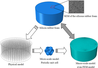
Gamma radiation induced compressive response of silicon rubber foam: Experiments and modeling
-
- Journal:
- Journal of Materials Research / Volume 34 / Issue 13 / 15 July 2019
- Published online by Cambridge University Press:
- 07 March 2019, pp. 2194-2200
- Print publication:
- 15 July 2019
-
- Article
- Export citation

Fabrication and characterization of microencapsulated n-octadecane with silk fibroin–silver nanoparticles shell for thermal regulation
-
- Journal:
- Journal of Materials Research / Volume 34 / Issue 12 / 28 June 2019
- Published online by Cambridge University Press:
- 07 March 2019, pp. 2047-2056
- Print publication:
- 28 June 2019
-
- Article
- Export citation
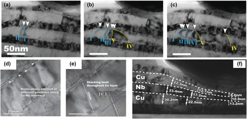
An overview of interface-dominated deformation mechanisms in metallic nanocomposites elucidated using in situ straining in a TEM
-
- Journal:
- Journal of Materials Research / Volume 34 / Issue 9 / 14 May 2019
- Published online by Cambridge University Press:
- 07 March 2019, pp. 1469-1478
- Print publication:
- 14 May 2019
-
- Article
- Export citation
Metal Organic Framework papers are a select set of papers in this issue
-
- Journal:
- Powder Diffraction / Volume 34 / Issue 1 / March 2019
- Published online by Cambridge University Press:
- 07 March 2019, p. 1
-
- Article
-
- You have access
- HTML
- Export citation
First-principles study of carbon capture and storage properties of porous MnO2 octahedral molecular sieve OMS-5
-
- Journal:
- Powder Diffraction / Volume 34 / Issue 1 / March 2019
- Published online by Cambridge University Press:
- 07 March 2019, pp. 13-20
-
- Article
- Export citation
Calendar of short courses and workshops
-
- Journal:
- Powder Diffraction / Volume 34 / Issue 1 / March 2019
- Published online by Cambridge University Press:
- 07 March 2019, pp. 85-86
-
- Article
-
- You have access
- HTML
- Export citation
67th Annual Denver X-ray Conference Report
-
- Journal:
- Powder Diffraction / Volume 34 / Issue 1 / March 2019
- Published online by Cambridge University Press:
- 07 March 2019, pp. 76-78
-
- Article
- Export citation
PDJ volume 34 issue 1 Cover and Back matter
-
- Journal:
- Powder Diffraction / Volume 34 / Issue 1 / March 2019
- Published online by Cambridge University Press:
- 07 March 2019, pp. b1-b6
-
- Article
-
- You have access
- Export citation

Characterization of dynamic and quasistatic compressive mechanical properties of ice-templated alumina–epoxy composites
-
- Journal:
- Journal of Materials Research / Volume 34 / Issue 6 / 28 March 2019
- Published online by Cambridge University Press:
- 07 March 2019, pp. 959-971
- Print publication:
- 28 March 2019
-
- Article
- Export citation
Calendar of forthcoming meetings
-
- Journal:
- Powder Diffraction / Volume 34 / Issue 1 / March 2019
- Published online by Cambridge University Press:
- 07 March 2019, pp. 87-88
-
- Article
-
- You have access
- HTML
- Export citation
