Refine search
Actions for selected content:
106116 results in Materials Science
MRS MOVERS & SHAKERS: Spread the good news!
-
- Journal:
- MRS Bulletin / Volume 45 / Issue 1 / January 2020
- Published online by Cambridge University Press:
- 10 January 2020, p. 72
- Print publication:
- January 2020
-
- Article
-
- You have access
- HTML
- Export citation
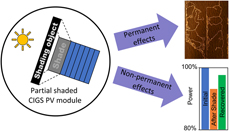
Reliability implications of partial shading on CIGS photovoltaic devices: A literature review
-
- Journal:
- Journal of Materials Research / Volume 34 / Issue 24 / 30 December 2019
- Published online by Cambridge University Press:
- 30 December 2019, pp. 3977-3987
- Print publication:
- 30 December 2019
-
- Article
- Export citation
JMR volume 34 issue 24 Cover and Front matter
-
- Journal:
- Journal of Materials Research / Volume 34 / Issue 24 / 30 December 2019
- Published online by Cambridge University Press:
- 30 December 2019, pp. f1-f5
- Print publication:
- 30 December 2019
-
- Article
-
- You have access
- Export citation
JMR volume 34 issue 24 Cover and Back matter
-
- Journal:
- Journal of Materials Research / Volume 34 / Issue 24 / 30 December 2019
- Published online by Cambridge University Press:
- 30 December 2019, pp. b1-b7
- Print publication:
- 30 December 2019
-
- Article
-
- You have access
- Export citation
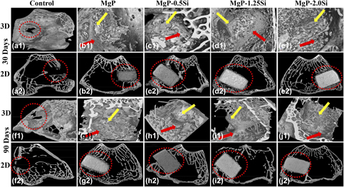
Quantitative assessment of degradation, cytocompatibility, and in vivo bone regeneration of silicon-incorporated magnesium phosphate bioceramics
-
- Journal:
- Journal of Materials Research / Volume 34 / Issue 24 / 30 December 2019
- Published online by Cambridge University Press:
- 30 December 2019, pp. 4024-4036
- Print publication:
- 30 December 2019
-
- Article
- Export citation
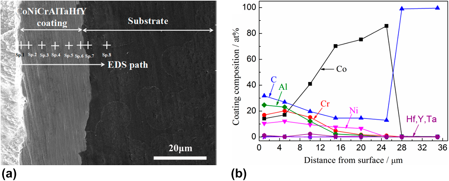
Improved oxidation resistance of CoNiCrAlTaHfY/Co coating on C/C composites by vapor phase surface alloying
-
- Journal:
- Journal of Materials Research / Volume 35 / Issue 5 / 16 March 2020
- Published online by Cambridge University Press:
- 23 December 2019, pp. 500-507
- Print publication:
- 16 March 2020
-
- Article
- Export citation

Planet–satellite nanostructures from inorganic nanoparticles: from synthesis to emerging applications
-
- Journal:
- MRS Communications / Volume 10 / Issue 1 / March 2020
- Published online by Cambridge University Press:
- 20 December 2019, pp. 112-122
- Print publication:
- March 2020
-
- Article
- Export citation

A perspective on overcoming water-related stability challenges in molecular and hybrid semiconductors
-
- Journal:
- MRS Communications / Volume 10 / Issue 1 / March 2020
- Published online by Cambridge University Press:
- 20 December 2019, pp. 98-111
- Print publication:
- March 2020
-
- Article
-
- You have access
- Open access
- HTML
- Export citation

Enhanced light-matter interactions in size tunable graphene–gold nanomesh
-
- Journal:
- MRS Communications / Volume 10 / Issue 1 / March 2020
- Published online by Cambridge University Press:
- 20 December 2019, pp. 135-140
- Print publication:
- March 2020
-
- Article
- Export citation
References
-
- Book:
- X-ray Microscopy
- Published online:
- 28 October 2019
- Print publication:
- 19 December 2019, pp 519-572
-
- Chapter
- Export citation
4 - Imaging Physics
-
- Book:
- X-ray Microscopy
- Published online:
- 28 October 2019
- Print publication:
- 19 December 2019, pp 71-198
-
- Chapter
- Export citation
Frontmatter
-
- Book:
- X-ray Microscopy
- Published online:
- 28 October 2019
- Print publication:
- 19 December 2019, pp i-iv
-
- Chapter
- Export citation
11 - Radiation Damage and Cryo Microscopy
-
- Book:
- X-ray Microscopy
- Published online:
- 28 October 2019
- Print publication:
- 19 December 2019, pp 457-495
-
- Chapter
- Export citation
Index
-
- Book:
- X-ray Microscopy
- Published online:
- 28 October 2019
- Print publication:
- 19 December 2019, pp 573-579
-
- Chapter
- Export citation
12 - Applications, and Future Prospects
-
- Book:
- X-ray Microscopy
- Published online:
- 28 October 2019
- Print publication:
- 19 December 2019, pp 496-514
-
- Chapter
- Export citation
Foreword
-
- Book:
- X-ray Microscopy
- Published online:
- 28 October 2019
- Print publication:
- 19 December 2019, pp xiii-xiv
-
- Chapter
- Export citation
Contents
-
- Book:
- X-ray Microscopy
- Published online:
- 28 October 2019
- Print publication:
- 19 December 2019, pp v-xi
-
- Chapter
- Export citation
6 - X-Ray Microscope Systems
-
- Book:
- X-ray Microscopy
- Published online:
- 28 October 2019
- Print publication:
- 19 December 2019, pp 241-258
-
- Chapter
- Export citation
7 - X-Ray Microscope Instrumentation
-
- Book:
- X-ray Microscopy
- Published online:
- 28 October 2019
- Print publication:
- 19 December 2019, pp 259-320
-
- Chapter
- Export citation
2 - A Bit of History
-
- Book:
- X-ray Microscopy
- Published online:
- 28 October 2019
- Print publication:
- 19 December 2019, pp 5-22
-
- Chapter
- Export citation
