Refine search
Actions for selected content:
106116 results in Materials Science
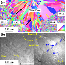
Dissimilar welding of Al0.1CoCrFeNi high-entropy alloy and AISI304 stainless steel
-
- Journal:
- Journal of Materials Research / Volume 34 / Issue 15 / 14 August 2019
- Published online by Cambridge University Press:
- 04 June 2019, pp. 2683-2694
- Print publication:
- 14 August 2019
-
- Article
- Export citation

Thermal desulfurization of pyrite: An in situ high-T neutron diffraction and DTA–TGA study
-
- Journal:
- Journal of Materials Research / Volume 34 / Issue 19 / 14 October 2019
- Published online by Cambridge University Press:
- 04 June 2019, pp. 3243-3253
- Print publication:
- 14 October 2019
-
- Article
- Export citation
-
To study thermal desulfurization of pyrite (FeS2), we conducted in situ neutron diffraction experiments in the temperature range 298–1073 K. On heating, pyrite remained stable up to 773 K, at which it started to decompose into pyrrhotite (Fe1−xS) and S2 gas. Rietveld analysis of the neutron data from 298 to 773 K allowed determination of the thermal expansion coefficient of pyrite (space group Pa $\bar 3$) to be αV = 3.7456 × 10⁻⁵ K−1, which largely results from the expansion of the Fe–S bond. With further increase in temperature to 1073 K, all the pyrite transformed to pyrrhotite (Fe1−xS) at 873 K. Unit-cell parameters of Fe1−xS (space group P63/mmc) increase on heating and decrease on cooling. However, the rates in cell expansion are larger than those in contraction. This hysteresis behavior can be attributed to continuous desulfurization of pyrrhotite (i.e., x in Fe1−xS decreases) with increasing temperature until the stoichiometric troilite (FeS) was formed at 1073 K. On cooling, troilite underwent a magnetic transition to an orthorhombic structure (space group Pnma) between 473 and 573 K. In addition, using differential thermal analysis (DTA) and thermogravimetric analysis (TGA) implemented with a differential scanning calorimeter, we performed kinetic measurements of pyrite decomposition. Detailed peak profile and Arrhenius (k = A exp(−Ea/RT)) analyses yielded an activation energy Ea of 302.3 ± 28.6 kJ/mol (based on DTA data) or 302.5 ± 26.4 kJ/mol (based on TGA data) and a ln(A) of 35.3 ± 0.1.

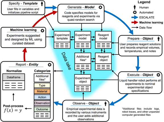
Experiment Specification, Capture and Laboratory Automation Technology (ESCALATE): a software pipeline for automated chemical experimentation and data management
-
- Journal:
- MRS Communications / Volume 9 / Issue 3 / September 2019
- Published online by Cambridge University Press:
- 04 June 2019, pp. 846-859
- Print publication:
- September 2019
-
- Article
- Export citation
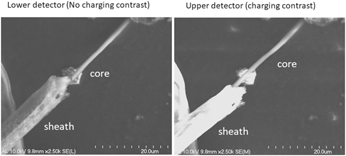
Formation of aligned core/sheath microfiber scaffolds with a poly-L-lactic acid (PLLA) sheath and a conductive poly(3,4-ethylenedioxythiophene) (PEDOT) core
-
- Journal:
- Journal of Materials Research / Volume 34 / Issue 11 / 14 June 2019
- Published online by Cambridge University Press:
- 04 June 2019, pp. 1931-1943
- Print publication:
- 14 June 2019
-
- Article
- Export citation

Atmospheric pressure high-power impulse plasma source for deposition of metallic coatings
-
- Journal:
- Journal of Materials Research / Volume 34 / Issue 12 / 28 June 2019
- Published online by Cambridge University Press:
- 04 June 2019, pp. 2078-2085
- Print publication:
- 28 June 2019
-
- Article
- Export citation

Effect of additions of metal submicron particles on properties of alumina matrix composites
-
- Journal:
- Journal of Materials Research / Volume 34 / Issue 17 / 16 September 2019
- Published online by Cambridge University Press:
- 04 June 2019, pp. 2983-2989
- Print publication:
- 16 September 2019
-
- Article
- Export citation
Fifty Materials That Make the World by Ian Baker
-
- Journal:
- MRS Bulletin / Volume 44 / Issue 6 / June 2019
- Published online by Cambridge University Press:
- 11 June 2019, p. 510
- Print publication:
- June 2019
-
- Article
-
- You have access
- HTML
- Export citation

New techniques for imaging and identifying defects in electron microscopy
-
- Journal:
- MRS Bulletin / Volume 44 / Issue 6 / June 2019
- Published online by Cambridge University Press:
- 11 June 2019, pp. 450-458
- Print publication:
- June 2019
-
- Article
- Export citation
ZnO Thin-Film Transistors for Cost-Efficient Flexible Electronics by Fábio Fedrizzi Vidor, Gilson Inácio Wirth, and Ulrich Hilleringmann
-
- Journal:
- MRS Bulletin / Volume 44 / Issue 6 / June 2019
- Published online by Cambridge University Press:
- 11 June 2019, p. 511
- Print publication:
- June 2019
-
- Article
-
- You have access
- HTML
- Export citation
From lab to market: Strategies to nanotechnology commercialization in Africa
-
- Journal:
- MRS Bulletin / Volume 44 / Issue 6 / June 2019
- Published online by Cambridge University Press:
- 11 June 2019, pp. 421-422
- Print publication:
- June 2019
-
- Article
-
- You have access
- HTML
- Export citation
Measurement of local strain
-
- Journal:
- MRS Bulletin / Volume 44 / Issue 6 / June 2019
- Published online by Cambridge University Press:
- 11 June 2019, pp. 459-464
- Print publication:
- June 2019
-
- Article
- Export citation
MRC volume 9 issue 2 Cover and Front matter
-
- Journal:
- MRS Communications / Volume 9 / Issue 2 / June 2019
- Published online by Cambridge University Press:
- 01 July 2019, pp. f1-f6
- Print publication:
- June 2019
-
- Article
-
- You have access
- Export citation

Advances in in situ nanomechanical testing
-
- Journal:
- MRS Bulletin / Volume 44 / Issue 6 / June 2019
- Published online by Cambridge University Press:
- 11 June 2019, pp. 438-442
- Print publication:
- June 2019
-
- Article
-
- You have access
- HTML
- Export citation
Reconstituting and protecting our oceans
-
- Journal:
- MRS Bulletin / Volume 44 / Issue 6 / June 2019
- Published online by Cambridge University Press:
- 11 June 2019, p. 433
- Print publication:
- June 2019
-
- Article
-
- You have access
- HTML
- Export citation
Materials Engineering: Bonding, Structure, and Structure-Property Relationships by Susan Trolier-McKinstry and Robert E. Newnham
-
- Journal:
- MRS Bulletin / Volume 44 / Issue 6 / June 2019
- Published online by Cambridge University Press:
- 11 June 2019, pp. 510-511
- Print publication:
- June 2019
-
- Article
-
- You have access
- HTML
- Export citation
South Africa and China begin student exchange program
-
- Journal:
- MRS Bulletin / Volume 44 / Issue 6 / June 2019
- Published online by Cambridge University Press:
- 11 June 2019, p. 429
- Print publication:
- June 2019
-
- Article
-
- You have access
- HTML
- Export citation

Impact of in situ nanomechanics on physical metallurgy
-
- Journal:
- MRS Bulletin / Volume 44 / Issue 6 / June 2019
- Published online by Cambridge University Press:
- 11 June 2019, pp. 465-470
- Print publication:
- June 2019
-
- Article
- Export citation
UK and European materials researchers concerned about Brexit
-
- Journal:
- MRS Bulletin / Volume 44 / Issue 6 / June 2019
- Published online by Cambridge University Press:
- 11 June 2019, pp. 428-429
- Print publication:
- June 2019
-
- Article
-
- You have access
- HTML
- Export citation
MRC volume 9 issue 2 Cover and Back matter
-
- Journal:
- MRS Communications / Volume 9 / Issue 2 / June 2019
- Published online by Cambridge University Press:
- 01 July 2019, pp. b1-b2
- Print publication:
- June 2019
-
- Article
-
- You have access
- Export citation
Clinical significance of three-dimensional printed biomaterials and biomedical devices
-
- Journal:
- MRS Bulletin / Volume 44 / Issue 6 / June 2019
- Published online by Cambridge University Press:
- 11 June 2019, pp. 494-504
- Print publication:
- June 2019
-
- Article
-
- You have access
- HTML
- Export citation
