We use cookies to distinguish you from other users and to provide you with a better experience on our websites. Close this message to accept cookies or find out how to manage your cookie settings.
To save content items to your account,
please confirm that you agree to abide by our usage policies.
If this is the first time you use this feature, you will be asked to authorise Cambridge Core to connect with your account.
Find out more about saving content to .
To save content items to your Kindle, first ensure no-reply@cambridge.org
is added to your Approved Personal Document E-mail List under your Personal Document Settings
on the Manage Your Content and Devices page of your Amazon account. Then enter the ‘name’ part
of your Kindle email address below.
Find out more about saving to your Kindle.
Note you can select to save to either the @free.kindle.com or @kindle.com variations.
‘@free.kindle.com’ emails are free but can only be saved to your device when it is connected to wi-fi.
‘@kindle.com’ emails can be delivered even when you are not connected to wi-fi, but note that service fees apply.
Tremor, which is defined as an oscillatory and rhythmic movement of a body part, is the most common movement disorder worldwide. The most frequent tremor syndromes are tremor in Parkinson’s disease, essential tremor, and dystonic tremor syndromes, whereas Holmes tremor, orthostatic tremor, and palatal tremor are less common in clinical practice. The pathophysiology of tremor consists of enhanced oscillatory activity in brain circuits, which are ofen modulated by tremor-related afferent signals from the periphery. The cerebello-thalamo-cortical circuit and the basal ganglia play a key role in most neurologic tremor disorders, but with different roles in each disorder. Here we review the pathophysiology of tremor, focusing both on neuronal mechanisms that promote oscillations (automaticity and synchrony) and circuit-level mechanisms that drive and maintain pathologic oscillations.
The neural correlates underlying late-life depressive symptoms and cognitive deterioration are largely unclear, and little is known about the role of chronic physical conditions in such association. This research explores both concurrent and longitudinal associations between late-life depressive symptoms and cognitive functions, with examining the neural substrate and chronic vascular diseases (CVDs) in these associations.
Methods
A total of 4109 participants (mean age = 65.4, 63.0% females) were evaluated for cognitive functions through various neuropsychological assessments. Depressive symptoms were assessed by the Geriatric Depression Scale and CVDs were self-reported. T1-weighted magnetic resonance imaging (MRI), diffusion tensor imaging, and functional MRI (fMRI) data were acquired in a subsample (n = 791).
Results
Cognitively, higher depressive symptoms were correlated with poor performance across all cognitive domains, with the strongest association with episodic memory (r = ‒0.138, p < 0.001). Regarding brain structure, depressive symptoms were negatively correlated with thalamic volume and white matter integrity. Further, white matter integrity was found to mediate the longitudinal association between depressive symptoms and episodic memory (indirect effect = −0.017, 95% CI −0.045 to −0.002) and this mediation was only significant for those with severe CVDs (β = −0.177, p = 0.008).
Conclusions
This study is one of the first to provide neural evidence elucidating the longitudinal associations between late-life depressive symptoms and cognitive dysfunction. Additionally, the severity of CVDs strengthened these associations, which enlightens the potential of managing CVDs as an intervention target for preventing depressive symptoms-related cognitive decline.
Major depressive disorder (MDD) is characterized by deficient reward functions in the brain. However, existing findings on functional alterations during reward anticipation, reward processing, and learning among MDD patients are inconsistent, and it was unclear whether a common reward system implicated in multiple reward functions is altered in MDD. Here we meta-analyzed 18 past studies that compared brain reward functions between adult MDD patients (N = 477, mean age = 26.50 years, female = 59.40%) and healthy controls (N = 506, mean age = 28.11 years, females = 55.58%), and particularly examined group differences across multiple reward functions. Jack-knife sensitivity and subgroup meta-analyses were conducted to test robustness of findings across patient comorbidity, task paradigm, and reward nature. Meta-regression analyses assessed the moderating effect of patient symptom severity and anhedonia scores. We found during reward anticipation, MDD patients showed lower activities in the lateral prefrontal-thalamus circuitry. During reward processing, patients displayed reduced activities in the right striatum and prefrontal cortex, but increased activities in the left temporal cortex. During reward learning, patients showed reduced activity in the lateral prefrontal–thalamic–striatal circuitry and the right parahippocampal–occipital circuitry but higher activities in bilateral cerebellum and the left visual cortex. MDD patients showed decreased activity in the right thalamus during both reward anticipation and learning, and in the right caudate during both reward processing and learning. Larger functional changes in MDD were observed among patients with more severe symptoms and higher anhedonia levels. The thalamic-striatal circuitry functional alterations could be the key neural mechanism underlying MDD patients overarching reward function deficiencies.
Essential tremor (ET) is the most common movement disorder, characterized by bilateral action tremors of the upper extremities. Surgical interventions can be considered for severe cases that are refractory to medication. Magnetic resonance-guided focused ultrasound (MRgFUS) of the ventral intermediate nucleus of the thalamus (Vim) is a recently approved, minimally invasive treatment for unilateral tremor. While patients are generally pleased with unilateral treatment, many patients are bothered by tremor on the untreated side. Historically, bilateral thalamotomy has been associated with a higher rate of adverse events, including cognitive impairment. MRgFUS Vim thalamotomy for bilateral tremor is currently being investigated. The goal of the present study was to evaluate the effect of bilateral MRgFUS Vim thalamotomy on cognition.
Participants and Methods:
Twelve patients with medication-refractory essential tremor (mean age = 68.77 +/- 11.78 years, mean education = 14.34 +/- 2.71 years, 8 male) were included in the present study. Three of the 12 patients met criteria for mild cognitive impairment (MCI). All patients successfully underwent unilateral MRgFUS thalamotomy at least 48 weeks before the second thalamotomy. A battery of neuropsychological tests was administered to patients before (considered baseline in the present study) and three months following the second thalamotomy. Baseline evaluations occurred on average 144.64 +/- 91.53 weeks (range: 55.00 - 346.58) after the first thalamotomy. The neuropsychological battery assessed domains of processing speed (Oral Symbol Digit Modalities Test, D-KEFS Color-Word Naming and Reading), attention (WAIS-IV Digit Span Forward), executive function (D-KEFS Color-Word Inhibition and Inhibition/Switching), working memory (WAIS-IV Digit Span Backward and Sequencing), verbal fluency (D-KEFS Letter Fluency and Animal Fluency), confrontation naming (Boston Naming Test), verbal memory (Hopkins Verbal Learning Test-Revised), and visuospatial perception (Judgment of Line Orientation). Alternate versions of tests were used when possible. Cognitive changes were analyzed at the group and individual level. Group level changes were assessed with paired sample t-tests (corrected for multiple comparisons). At the individual level, postoperative declines > 1.5 SD from baseline were considered clinically significant.
Results:
Participants’ baseline intellectual functioning ranged from low average to superior (as measured by the WTAR). The mean baseline score on the Montreal Cognitive Assessment was 24.58 (range: 17 - 30). At the group level, there were no significant changes in cognitive scores from baseline to follow-up (all p values > 0.635). At the individual level, one patient with MCI declined > 1.5 SD on the verbal memory composite. No other patients showed declines > 1.5 SD.
Conclusions:
Our preliminary findings suggest that bilateral MRgFUS Vim thalamotomy is relatively safe from a cognitive perspective. However, a single patient with MCI exhibited clinically significant postoperative decline in verbal memory. Future studies with larger sample sizes are needed to investigate the factors that increase the risk of postoperative cognitive decline, including pre-existing cognitive impairment, older age, and lesion size.
The ability to extinguish a maladaptive conditioned fear response is crucial for healthy emotional processing and resiliency to aversive experiences. Therefore, enhancing fear extinction learning has immense potential emotional and health benefits. Mindfulness training enhances both fear conditioning and recall of extinguished fear; however, its effects on fear extinction learning are unknown. Here we investigated the impact of mindfulness training on brain mechanisms associated with fear-extinction learning, compared to an exercise-based program.
Methods
We investigated BOLD activations in response to a previously learned fear-inducing cue during an extinction paradigm, before and after an 8-week mindfulness-based stress reduction program (MBSR, n = 49) or exercise-based stress management education program (n = 27).
Results
The groups exhibited similar reductions in stress, but the MBSR group was uniquely associated with enhanced activation of salience network nodes and increased hippocampal engagement.
Conclusions
Our results suggest that mindfulness training increases attention to anticipatory aversive stimuli, which in turn facilitates decreased aversive subjective responses and enhanced reappraisal of the memory.
The motor thalamus is interconnected with the brainstem, cortex, and basal ganglia and plays major roles in planning, sequencing, and executing action. In this chapter, I highlight roles of input-defined thalamic circuits in motor sequence production and learning. Brainstem–motor thalamic pathways carry efference copy signals important for the production of both innate and learned motor sequences, for example, during saccades, grooming, and birdsong. Basal ganglia thalamocortical loops implement aspects of reinforcement learning, including the generation of motor exploration during vocal babbling. Classic "gating" models of basal ganglia–thalamic transmission fail to explain thalamic discharge during behavior, which instead appears strongly driven by cortical inputs. A challenge going forward is to determine if there are conserved principles of thalamic function across diverse motor thalamic subregions.
Cognitive control refers to our ability to regulate thoughts and actions for adaptive, goal-directed behaviors. Traditionally, cognitive control is thought to be mediated by the prefrontal cortex; however, the thalamus likely plays an important yet underappreciated role. This chapter reviews the role of the human thalamus in cognitive control. We first review anatomical, human functional neuroimaging, and human neuropsychology findings that have investigated the role of the human thalamus in two cognitive control functions: working memory and top-down biasing. To understand how the human thalamus mechanistically supports cognitive control, we then summarize operational principles of thalamocortical circuits from anatomical and neurophysiological studies. Finally, we present an overarching conceptual framework to describe how thalamocortical circuits implement different components of information processing necessary for cognitive control. In conclusion, we refute the traditional view that the thalamus passively relays signals to the cortex for purposeful processing. Instead, emerging evidence suggests that the thalamus actively modulates cortical activity and cortical network interactions to shape and coordinate information processes underlying cognitive control.
The cortex and thalamus are richly interconnected by feedforward and feedback pathways. For vision, the lateral geniculate nucleus (LGN) of the dorsal thalamus supplies the primary visual cortex with synaptically strong feedforward input that carries information about the external environment. The cortex, in turn, sends an even greater number of axons back to the LGN; however, these corticogeniculate inputs are relatively weak. Based on this anatomy, the cortex appears to be able to modulate the nature of the signals it receives from the LGN, potentially to meet the ongoing processing needs of the cortex. This chapter examines the relationship between the feedforward and feedback pathways interconnecting the LGN and visual cortex, with an emphasis on their organization with respect to the parallel processing streams originating in the retina. It also explores the influence of corticogeniculate feedback on vision.
Higher thalamic volume has been found in children with obsessive-compulsive disorder (OCD) and children with clinical-level symptoms within the general population (Boedhoe et al. 2017, Weeland et al. 2021a). Functionally distinct thalamic nuclei are an integral part of OCD-relevant brain circuitry.
Objectives
We aimed to study the thalamic nuclei volume in relation to subclinical and clinical OCD across different age ranges. Understanding the role of thalamic nuclei and their associated circuits in pediatric OCD could lead towards treatment strategies specifically targeting these circuits.
Methods
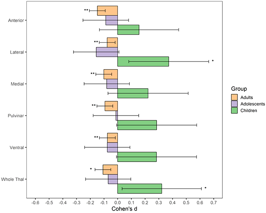
We studied the relationship between thalamic nuclei and obsessive-compulsive symptoms (OCS) in a large sample of school-aged children from the Generation R Study (N = 2500) (Weeland et al. 2021b). Using the data from the ENIGMA-OCD working group we conducted mega-analyses to study thalamic subregional volume in OCD across the lifespan in 2,649 OCD patients and 2,774 healthy controls across 29 sites (Weeland et al. 2021c). Thalamic nuclei were grouped into five subregions: anterior, ventral, intralaminar/medial, lateral and pulvinar (Figure 1).
Results
Both children with subclinical and clinical OCD compared with controls show increased volume across multiple thalamic subregions. Adult OCD patients have decreased volume across all subregions (Figure 2), which was mostly driven by medicated and adult-onset patients.
Conclusions
Our results suggests that OCD-related thalamic volume differences are global and not driven by particular subregions and that the direction of effects are driven by both age and medication status.
Sensory information enters the cerebral cortex through separate thalamocortical pathways that originate in different senses. One of these pathways links the dorsal lateral geniculate nucleus of the thalamus to the primary visual cortex and is crucial for mammalian vision. Over the past decades, there has been tremendous progress in understanding its functional organization, and new tools are allowing us to isolate, with increasing precision, its different components. Just as different senses remain segregated on their way to the cerebral cortex, the different properties of the visual stimulus also reach the primary visual cortex through separate geniculocortical pathways. On the one hand, these separate pathways underlie the parallel processing of stimulus position, eye of origin, light–dark polarity, and temporal dynamics, a strategy that is well preserved across species. On the other hand, the convergence of the different geniculocortical pathways in the visual cortex enables cortical neurons to extract features of the visual world that are not encoded by any geniculocortical pathway individually. This chapter reviews the current knowledge on the functional organization of this prominent thalamocortical pathway and concludes by raising key questions to be addressed in the future.
Survivors of patients with artery of Percheron infarction (API) often have a prolonged and disabling form of cognitive impairment that remains insufficiently characterized. We aimed to examine the clinical and cognitive features of API in the short and long term after stroke.
Methods:
We reviewed 6400 patients with a first-ever stroke included in the Stroke Registry between 2011 and 2021. The diagnosis of API was based on clinical diagnosis and imaging confirmation. All patients underwent neuropsychological assessment at hospital stay and 1 year after stroke. A z-score of each patients’ cognitive test point was calculated, and a z-score inferior to 2 was considered as pathological.
Results:
Of the 10 patients enrolled, all had cognitive impairment, consciousness, and behavioral disorders at stroke onset. Six patients had pure bilateral thalamic involvement while four had bilateral thalamic and rostral midbrain involvement. At 12 months, 50% of patients had global mental state scores 2 SD below the population mean (z-score mean ± SD, −2.17 ± 0.4). Most of the prefrontal cortex cognitive processes including executive functions such as planning and cognitive control (z-score mean ± SD, −3.92 ± 0.3), processing speed (−4.42 ± 0.5), working memory (−3.97 ± 0.3) were severely impaired at stroke onset. Especially in patients with thalamic and rostral midbrain involvement, deficiencies in executive function (z-score mean ± SD, −2.60 ± 0.4), processing speed (−2.22 ± 0.5), working (−3.76 ± 0.4), and episodic memory (−2.23 ± 0.3) continued 12 months after stroke.
Conclusions:
The occlusion of the artery of Percheron results in severe behavioral and cognitive disorders in the short and long term after stroke.
The context-based selection of semantic representations is presented as an essential issue in understanding the interpretation of speech meaning. Clinical findings serve to illustrate the role of the thalamus, the basal ganglia, and the cerebellum in processing subtle context-related differences in meaning. The paucity of findings relating to spoken language is emphasized along with the need to specify neuropragmatic principles of context-based activations of semantic and episodic representations.With a view on developing such principles, several relevant findings are reviewed relating to cortico-thalamic interactions and the pivotal role of the motor thalamus in integrating multisensory information from basal-ganglia circuits and the cerebellum. The on-line selection of semantic and episodic representations is also discussed in terms of experiments on the role of the hippocampus and frontal circuits suggesting some parallels with navigation, but the on-line processing of speech requires a chunking of action-related sequences which appears to involve the basal ganglia and critical cortico-thalamic loops.
Childhood trauma is a vulnerability factor for the development of obsessive–compulsive disorder (OCD). Empirical findings suggest that trauma-related alterations in brain networks, especially in thalamus-related regions, have been observed in OCD patients. However, the relationship between childhood trauma and thalamic connectivity in patients with OCD remains unclear. The present study aimed to examine the impact of childhood trauma on thalamic functional connectivity in OCD patients.
Methods
Magnetic resonance imaging resting-state scans were acquired in 79 patients with OCD, including 22 patients with a high level of childhood trauma (OCD_HCT), 57 patients with a low level of childhood trauma (OCD_LCT) and 47 healthy controls. Seven thalamic subdivisions were chosen as regions of interest (ROIs) to examine the group difference in thalamic ROIs and whole-brain resting-state functional connectivity (rsFC).
Results
We found significantly decreased caudate-thalamic rsFC in OCD patients as a whole group and also in OCD_LCT patients, compared with healthy controls. However, OCD_HCT patients exhibited increased thalamic rsFC with the prefrontal cortex when compared with both OCD_LCT patients and healthy controls.
Conclusions
Taken together, OCD patients with high and low levels of childhood trauma exhibit different pathological alterations in thalamic rsFC, suggesting that childhood trauma may be a predisposing factor for some OCD patients.
To describe the experience with Anterior Nucleus of the Thalamus-Deep Brain Stimulation (ANT-DBS) for the treatment of epilepsy at a Canadian Center.
Methods:
All patients who underwent ANT-DBS implantation between 2013 (first patient implanted at our center) and 2020 were included. These patients had therapy-resistant epilepsy (TRE), were not candidates for resective surgery, and failed vagus nerve stimulation (VNS) treatment. Baseline of monthly seizure frequency was calculated within 3 months prior to VNS placement. Monthly seizure frequency was assessed at different points along the timeline: 3 months before ANT-DBS implantation as well as 3, 6, 12, 24, 36, 48, 60, and 72 months after ANT-DBS device placement. At each time point, seizure frequency was compared to baseline.
Results:
Six patients were implanted with ANT-DBS. Three (50%) patients had multifocal epilepsy, one (16.6%) had focal epilepsy, and two (33.4%) had combined generalized and focal epilepsy. Two patients with multifocal epilepsy experienced a seizure reduction >50% in the long-term follow-up. Three (50%) patients did not showed improvement: two with combined generalized and focal epilepsy and one with focal epilepsy. There were not surgical or device-related side effects. Two (33.3%) patients presented mild and transient headaches as a stimulation-related side effect.
Conclusion:
ANT-DBS is an effective and safe treatment for focal TRE. Our experience suggests that patients with multifocal epilepsy due to regional lesion may benefit from ANT-DBS the most. Further investigations are required to determine optimal parameters of stimulation.
Social anxiety disorder (SAD) is characterized by anxiety regarding social situations, avoidance of external social stimuli, and negative self-beliefs. Virtual reality self-training (VRS) at home may be a good interim modality for reducing social fears before formal treatment. This study aimed to find neurobiological evidence for the therapeutic effect of VRS.
Methods
Fifty-two patients with SAD were randomly assigned to a VRS or waiting list (WL) group. The VRS group received an eight-session VRS program for 2 weeks, whereas the WL group received no intervention. Clinical assessments and functional magnetic resonance imaging scanning with the distress and speech evaluation tasks were repeatedly performed at baseline and after 3 weeks.
Results
The post-VRS assessment showed significantly decreased anxiety and avoidance scores, distress index, and negative evaluation index for ‘self’, but no change in the negative evaluation index for ‘other’. Patients showed significant responses to the distress task in various regions, including both sides of the prefrontal regions, occipital regions, insula, and thalamus, and to the speech evaluation task in the bilateral anterior cingulate cortex. Among these, significant neuronal changes after VRS were observed only in the right lingual gyrus and left thalamus.
Conclusions
VRS-induced improvements in the ability to pay attention to social stimuli without avoidance and even positively modulate emotional cues are based on functional changes in the visual cortices and thalamus. Based on these short-term neuronal changes, VRS can be a first intervention option for individuals with SAD who avoid society or are reluctant to receive formal treatment.
Schizophrenia (SZ) is a complex disorder characterized by a range of behavioral and cognitive symptoms as well as structural and functional alterations in multiple cortical and subcortical structures. SZ is associated with reduced functional network connectivity involving core regions such as the anterior cingulate cortex (ACC) and the thalamus. However, little is known whether effective coupling, the directed influence of one structure over the other, is altered during rest in the ACC–thalamus network.
Methods
We collected resting-state fMRI and diffusion-weighted MRI data from 18 patients and 20 healthy controls. We analyzed fronto-thalamic effective connectivity using dynamic causal modeling for cross-spectral densities in a network consisting of the ACC and the left and right medio-dorsal thalamic regions. We studied structural connectivity using fractional anisotropy (FA).
Results
We found decreased coupling strength from the right thalamus to the ACC and from the right thalamus to the left thalamus, as well as increased inhibitory intrinsic connectivity in the right thalamus in patients relative to controls. ACC-to-left thalamus coupling strength correlated with the Positive and Negative Syndrome Scale (PANSS) total positive syndrome score and with delusion score. Whole-brain structural analysis revealed several tracts with reduced FA in patients, with a maximum decrease in white matter tracts containing fronto-thalamic and cingulo-thalamic fibers.
Conclusions
We found altered effective and structural connectivity within the ACC–thalamus network in SZ. Our results indicate that ACC–thalamus network activity at rest is characterized by reduced thalamus-to-ACC coupling. We suggest that positive symptoms may arise as a consequence of compensatory measures to imbalanced fronto-thalamic coupling.
This study applied 1H-MRS in the thalamus of schizophrenic patients and healthy subjects.
There were no differences in the metabolite ratios (NAA/Cr, Cho/Cr or mI/Cr) between the two groups. Relationships were noted between NAA/Cr and age in patients with a trend toward this correlation in controls, suggesting an effect of age on the metabolism of the thalamus.
Poor response to dopaminergic antipsychotics constitutes a major challenge in the treatment of psychotic disorders and markers for non-response during first-episode are warranted. Previous studies have found increased levels of glutamate and γ-aminobutyric acid (GABA) in non-responding first-episode patients compared to responders, but it is unknown if non-responders can be identified using reference levels from healthy controls (HCs).
Methods
Thirty-nine antipsychotic-naïve patients with first-episode psychosis and 36 matched HCs underwent repeated assessments with the Positive and Negative Syndrome Scale and 3T magnetic resonance spectroscopy. Glutamate scaled to total creatine (/Cr) was measured in the anterior cingulate cortex (ACC) and left thalamus, and levels of GABA/Cr were measured in ACC. After 6 weeks, we re-examined 32 patients on aripiprazole monotherapy and 35 HCs, and after 26 weeks we re-examined 30 patients on naturalistic antipsychotic treatment and 32 HCs. The Andreasen criteria defined non-response.
Results
Before treatment, thalamic glutamate/Cr was higher in the whole group of patients but levels normalized after treatment. ACC levels of glutamate/Cr and GABA/Cr were lower at all assessments and unaffected by treatment. When compared with HCs, non-responders at week 6 (19 patients) and week 26 (16 patients) had higher baseline glutamate/Cr in the thalamus. Moreover, non-responders at 26 weeks had lower baseline GABA/Cr in ACC. Baseline levels in responders and HCs did not differ.
Conclusion
Glutamatergic and GABAergic abnormalities in antipsychotic-naïve patients appear driven by non-responders to antipsychotic treatment. If replicated, normative reference levels for glutamate and GABA may aid estimation of clinical prognosis in first-episode psychosis patients.