To save content items to your account,
please confirm that you agree to abide by our usage policies.
If this is the first time you use this feature, you will be asked to authorise Cambridge Core to connect with your account.
Find out more about saving content to .
To save content items to your Kindle, first ensure no-reply@cambridge.org
is added to your Approved Personal Document E-mail List under your Personal Document Settings
on the Manage Your Content and Devices page of your Amazon account. Then enter the ‘name’ part
of your Kindle email address below.
Find out more about saving to your Kindle.
Note you can select to save to either the @free.kindle.com or @kindle.com variations.
‘@free.kindle.com’ emails are free but can only be saved to your device when it is connected to wi-fi.
‘@kindle.com’ emails can be delivered even when you are not connected to wi-fi, but note that service fees apply.
This chapter’s purpose is to present the aim of the book, its rationale, focus, approach and the basic concepts necessary to make sense of what follows. The first part outlines the aim and approach, focussing on the impact of diet on the human brain and mind, alongside an outline of the content. The second part provides an overview of the core knowledge and methods that underpin research into diet, brain and mind. This starts with basic nutritional (energy needs, macronutrients, micronutrients) and physiological concepts (metabolism, digestion, regulation). It then covers the key issue of dietary measurement (self-report, observation, biomarkers, manipulation) and its limitations (accuracy, demand, stability over days and decades). The latter part examines the measurement of mind and brain – and its limitations – concentrating, respectively, on neuropsychological tests and imaging approaches. The final part describes our study inclusion criteria, and our rationale for favouring those with a whole diet focus.
This chapter examines the implications arising from the book’s content, and draws some general conclusions. Section 10.2 considers implications on a chapter-by-chapter basis, covering the small effect of breastfeeding on cognition, the nature of energy metabolism in the brain, dietary components versus patterns, the gut–brain axis, caffeine and sugar, immorality and hunger, lifespan extension and depression as a common consequence of nutritional deficiency. Section 10.3 presents conclusions and future directions, organised under three headings. First, the necessity to improve dietary recording methods, examining biomarkers for individual foods, active and passive food image collection, and monitoring eating. Second, nutraceutical and nutrigenomics, with the former’s failure so far to deliver concrete benefits – and why – and the latter’s potential to explain this through understanding individual differences. Third, the translation and reproducibility crises in biomedicine, and some consideration of their solutions as they apply here.
This chapter – focussing on adults – concerns the effects on brain and behaviour of deficiencies in vitamins, minerals and macronutrients, which cannot be synthesised in the body. Section 9.2 examines the neurobehavioural consequences of hypovitaminosis (intake below that recommended) and deficiency for each vitamin, including thiamine (Wernicke’s encephalopathy and Korsakoff’s syndrome), NAD (pellagra) and folate (depression). Section 9.3 covers mineral deficiencies, with notable impacts from iodine (hippocampal impairment and links to neurodegeneration in later life), selenium (hippocampal impairment) and zinc (depression). Section 9.4 examines the two essential macronutrient deficiencies. One covers omega-3 and omega-6 fatty acids, with deficiency linked to depression and neurodegeneration. The other covers the essential amino acids and the brain’s unique deficiency detection mechanism. Depression seems to be a common consequence of deficiency, and deficiency in mid-to-later life seems to link to neurodegeneration, but supplementation generally of individual micronutrients has not revealed much benefit in this regard.
Everybody eats, and what we eat – or do not – affects the brain and mind. There is significant general, applied, academic, and industry interest about nutrition and the brain, yet there is much misinformation and no single reliable guide. 
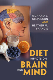
Diet Impacts on Brain and Mind provides a comprehensive account of this emerging multi-disciplinary science, exploring the acute and chronic impacts of human diet on the brain and mind. It has a primarily human focus and is broad in scope, covering wide-ranging topics like brain development, whole diets, specific nutrients, research methodology, and food as a drug. It is written in an accessible format and is of interest to undergraduate and graduate students studying nutritional neuroscience and related disciplines, healthcare professionals with an applied interest, industry researchers seeking topic overviews, and interested general readers.
The lampreys (Cyclostomes) represent the oldest group of now-living vertebrates that diverged from the vertebrate evolutionary line leading to mammals 560 million years ago. It is therefore of particular interest to consider if there is a thalamus similar to that of other vertebrates and examine how it is organised. The lamprey thalamus relays both visual and somatosensory information to the cortex (also called the pallium). In addition, the thalamus receives input from both the optic tectum (superior colliculus) and pretectum, as in other vertebrates, and there is, furthermore, a thalamic projection to the striatum from cells located in the periventricular area of the thalamus. Essentially, the basic compartments of the mammalian thalamus are thus represented in the lamprey but with a much smaller number of neurons. The implication is that the essential features of the thalamus had already evolved at the point when the cyclostome lineage diverged from that leading to other vertebrates. We review here what is known regarding the lamprey thalamus from the perspective of anatomy, transmitters, and neurophysiology and also how it compares to mammals, as well as other groups of vertebrates.
The medial geniculate body (MGB) of the thalamus plays a critical role in transforming the dense, high-fidelity auditory coding of the brainstem and midbrain to the sparse, abstract coding used throughout the forebrain to represent the perceptual qualities and behavioral meaning of sound. Here, we review the current state of knowledge on the connectivity, functional processing, and plasticity of interconnected neural circuits linking the MGB and the auditory cortex (ACtx). We describe new findings on the activation of corticothalamic neurons prior to expected sounds and specializations for encoding sound features that unfold on slow timescales that first emerge at the level of the MGB and ACtx. We review the literature on the development and plasticity of the MGB and ACtx, with a particular emphasis on how early auditory experience and adult learning modify sound processing at the level of thalamocortical synapses, circuits, and integrated neural systems. Despite its critical role as the root of forebrain sound processing, direct recordings from anatomically or genetically identified MGB cell types are rarely performed. We conclude by identifying several important knowns and unknowns about the distinct patterns of connectivity and functional specializations of the ventral, dorsal, and medial subdivisions of the MGB that await future investigation.
The motor thalamus is interconnected with the brainstem, cortex, and basal ganglia and plays major roles in planning, sequencing, and executing action. In this chapter, I highlight roles of input-defined thalamic circuits in motor sequence production and learning. Brainstem–motor thalamic pathways carry efference copy signals important for the production of both innate and learned motor sequences, for example, during saccades, grooming, and birdsong. Basal ganglia thalamocortical loops implement aspects of reinforcement learning, including the generation of motor exploration during vocal babbling. Classic "gating" models of basal ganglia–thalamic transmission fail to explain thalamic discharge during behavior, which instead appears strongly driven by cortical inputs. A challenge going forward is to determine if there are conserved principles of thalamic function across diverse motor thalamic subregions.
The thalamocortical system underlies sensory perception, brain-state regulation, movement execution, and cognition. The thalamus and cortex are generated from separate sectors of the embryonic forebrain, and their reciprocal axonal projections have to grow across a complex cellular terrain through it to innervate each other. The corticofugal and thalamocortical projections start to develop synchronously at very early stages when the distances are minimal. The initial topographical organization of these axons is guided by diencephalic and telencephalic molecular gradients. Transient axonal bundles and streams of migrating cell populations then act as a guiding scaffold for these projections. Once they reach their target, the thalamocortical fibers rearrange within the transient subplate zone and later innervate the cortical plate, whereas the corticofugal axons originate from layer 5, layer 6, and subplate/layer 6b neurons and follow a specific developmental sequence as they approach the thalamus, where some of them accumulate before they enter the first -and higher-order thalamic nuclei according to their subtypes and laminar origin. Both thalamocortical and corticothalamic projections are plastic and can reshape after alterations in sensory input or various lesions. Understanding the mechanisms underlying their development and remodeling is vital to comprehending the establishment and plasticity of cortical representations.
Cognitive control refers to our ability to regulate thoughts and actions for adaptive, goal-directed behaviors. Traditionally, cognitive control is thought to be mediated by the prefrontal cortex; however, the thalamus likely plays an important yet underappreciated role. This chapter reviews the role of the human thalamus in cognitive control. We first review anatomical, human functional neuroimaging, and human neuropsychology findings that have investigated the role of the human thalamus in two cognitive control functions: working memory and top-down biasing. To understand how the human thalamus mechanistically supports cognitive control, we then summarize operational principles of thalamocortical circuits from anatomical and neurophysiological studies. Finally, we present an overarching conceptual framework to describe how thalamocortical circuits implement different components of information processing necessary for cognitive control. In conclusion, we refute the traditional view that the thalamus passively relays signals to the cortex for purposeful processing. Instead, emerging evidence suggests that the thalamus actively modulates cortical activity and cortical network interactions to shape and coordinate information processes underlying cognitive control.
The architecture of the thalamus and its reciprocal connections with multiple cortical and subcortical structures are essential to the generation of the thalamo-cortical network oscillations associated with attention, sleep, and consciousness. This chapter provides an overview of the cellular mechanisms underlying thalamo-cortical network oscillations occurring during sleep and their contribution to the architecture of the sleep–wake cycle, including the onset and stability of non–rapid eye movement (NREM) and rapid eye movement (REM) sleep. It further summarizes the influence of the brainstem neuromodulatory system on thalamo-cortical network activity during wakefulness and sleep. Finally, the association between these mechanisms and synaptic plasticity in thalamo-cortical networks is described in the context of sleep-dependent consolidation, or weakening, of previously acquired information in health and disease.
The three anterior thalamic nuclei and the nucleus reuniens are essential for spatial navigation, yet their exact role in this function remains elusive. Specifically, it remains to be answered whether the thalamus acts a simple relay of spatial and executive signals or whether it critically operates on its inputs to convey processed signals to its cortical targets. The anterior thalamus and nucleus reuniens are at the center stage of anatomical networks that share one common aspect: their association with the hippocampus. Here, I review the large body of literature, starting from the classic Papez circuits, which describe how these thalamic nuclei are interconnected with subcortical, medial cortex, and parahippocampal regions, as well as their neuromodulatory inputs. I then provide an overview of the spatial and other electrophysiological correlates of anterior thalamic and reuniens neurons and of how their firing and oscillatory properties depend on ongoing behavior. Finally, I discuss the clinical and experimental evidence pointing to the role of the thalamus in navigation and, specifically, how spatial and executive signals are processed in thalamocortical loops. I conclude by discussing how the same thalamic circuits may be at play in the processing of episodic memories during sleep.
The thalamus traditionally is divided into the dorsal and ventral divisions, with both divisions divided into groups of nuclei. More recent approaches have defined divisions based on functional features, development, and/or evolutionary relationships. The thalamic nuclei are inextricably related to the corresponding divisions of the telencephalon, which is now recognized to have four developmental pallial sectors (the medial sector–derived hippocampal cortices; dorsal sector–derived neocortex; and lateral and ventral sector–derived claustrum, pallial amygdala, and related lateral nuclei in mammals) and the subpallium (which includes the septal nuclei, subpallial amygdala, and basal ganglia in mammals). The two evolutionary divisions of the dorsal thalamus, present in all jawed vertebrates, are the lemnothalamus and collothalamus. In amniotes, elaboration of different pallial sectors and their related dorsal thalamic nuclei was divergent, with the lemnothalamic-related, allocentric spatial mapping abilities selected for early in the line to mammals and the collothalamic-related, egocentric spatial mapping abilities selected for early in the line to reptiles, including birds. Secondarily, the collothalamic system was elaborated in mammals (e.g., LP/pulvinar in primates) and the lemnothalamic system in reptiles (e.g., primary visual and somatosensory nuclei in birds). Commonalities of thalamotelencephalic circuit motifs evolved in both lineages, supporting the functions of complex cognition and consciousness.
This chapter applies a perspective from biophysically grounded computational modeling to explore how the intrinsic properties of thalamic microcircuits support the computational roles that the thalamus plays in perceptual and cognitive functions. A key focus is on the modeling of neurophysiological activity in the thalamus as nonlinear dynamical systems. Dynamical modeling can give insight into thalamic function across levels of analysis, including cellular channel properties, synaptic plasticity, and anatomical connectivity. This chapter reviews how the interplay between cellular and circuit mechanisms supports thalamic contributions to neural oscillations, regulation of brain state, top-down attentional control of sensory processing, and other cognitive functions. Understanding circuit function through biophysically grounded computational modeling and dynamical systems perspectives can also provide insight into how cellular and synaptic alterations caused by pharmacology or disease can impair thalamic function.
The somatosensory thalamocortical system has proven a tractable model for dissecting how different neuronal populations sculpt bidirectional information exchange between the thalamus and the cortex. This chapter reviews corticothalamic (CT) pathways from layers 5 (L5) and 6 (L6) of the primary somatosensory (S1) cortex to first-order ventroposterior (VP) and higher-order posterior medial (POm) somatosensory thalamic nuclei. With a focus on insights gained from recent cell-type–specific approaches in rodent models, we contrast L5 and L6 CT pathways at the scales of network architecture, anatomical connectivity, and physiological characteristics. We further compare the distinct feedforward inhibitory circuits engaged by L6 and L5 CT pathways, which involve the thalamic reticular nucleus and extrathalamic inhibitory nuclei, respectively. Where data exist, we discuss short- and long-term synaptic dynamics of the specific CT circuits. We close with a discussion of the proposed functions of these distinct pathways in conveying “top-down” cortical signals for both the modulation of thalamic processing of sensory information and the transmission of information between cortical regions.
This chapter traces the scientific and clinical evidence supporting the key role of the central thalamus in forebrain arousal-regulation mechanisms. Beginning from the original studies of Moruzzi and Magoun in the late 1940s, the evolving concepts of defined pathways extending from the central regions of the thalamus that modulate arousal via their projections to cortical and striatal neuronal populations are carefully examined.Anatomical and physiological distinctions between the main subnuclei grouped within the often loosely defined "central thalamus," the more rostral central lateral and paracentral nuclei (CL/Pc), and the more caudal centromedian-parafasicularis complex (Cm-Pf) are reviewed across experimental studies in rodents, cats, and non-human primates. In addition, relevant human neuroimaging, neurophysiological, lesion correlation, and interventional neurosurgical studies are compared and discussed in the context of the reviewed basic neuroscience studies characterizing the central thalamus. Several scientific controversies surrounding the contributions of the central thalamus to forebrain arousal are specifically considered. This synoptic review is further used to unpack the rationale for the use of electrical stimulation centered on the CL/Pc populations to support impaired arousal regulation resulting from relatively deafferented frontal cortical and striatal populations in patients across a wide range of outcomes following coma induced by structural brain injuries.
The cortex and thalamus are richly interconnected by feedforward and feedback pathways. For vision, the lateral geniculate nucleus (LGN) of the dorsal thalamus supplies the primary visual cortex with synaptically strong feedforward input that carries information about the external environment. The cortex, in turn, sends an even greater number of axons back to the LGN; however, these corticogeniculate inputs are relatively weak. Based on this anatomy, the cortex appears to be able to modulate the nature of the signals it receives from the LGN, potentially to meet the ongoing processing needs of the cortex. This chapter examines the relationship between the feedforward and feedback pathways interconnecting the LGN and visual cortex, with an emphasis on their organization with respect to the parallel processing streams originating in the retina. It also explores the influence of corticogeniculate feedback on vision.