Skip to main content Accessibility help
×
  • Cited by 7
Publisher:
Cambridge University Press
Online publication date:
March 2013
Print publication year:
2013
Online ISBN:
9781139088268

Book description

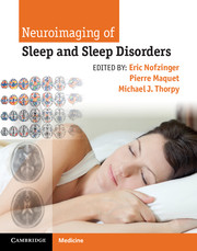
This up-to-date, superbly illustrated book is a practical guide to the effective use of neuroimaging in the patient with sleep disorders. There are detailed reviews of new neuroimaging techniques – including CT, MRI, advanced MR techniques, SPECT and PET – as well as image analysis methods, their roles and pitfalls. Neuroimaging of normal sleep and wake states is covered plus the role of neuroimaging in conjunction with tests of memory and how sleep influences memory consolidation. Each chapter carefully presents and analyzes the key findings in patients with sleep disorders indicating the clinical and imaging features of the various sleep disorders from clinical presentation to neuroimaging, aiding in establishing an accurate diagnosis. Written by neuroimaging experts from around the world, Neuroimaging of Sleep and Sleep Disorders is an invaluable resource for both researchers and clinicians including sleep specialists, neurologists, radiologists, psychiatrists, psychologists.

Reviews

“…Invaluable resource for researchers and clinicians in…sleep medicine, neurology, radiology, psychiatry, and psychology. A particular strength of the book is the incorporation of color figures and graphs of neuroimaging results.”

- Doody's Review Service

Refine List

Actions for selected content:

Select all | Deselect all
  • View selected items
  • Export citations
  • Download PDF (zip)
  • Save to Kindle
  • Save to Dropbox
  • Save to Google Drive

Save Search

You can save your searches here and later view and run them again in "My saved searches".

Please provide a title, maximum of 40 characters.
×

Contents


Page 3 of 3


  • Chapter 49 - Neuroimaging of sleep-related epilepsies
    pp 384-390
  • View abstract

    Summary

    This chapter focuses on classic congenital central hypoventilation syndrome (CCHS) cases in which symptoms appear early in life. The principal issues in CCHS are to determine what brain structures are damaged in CCHS to cause the loss of CO2 and O2 sensitivity, disturbances in autonomic function, and other affective and cognitive deficits. Among the processes affected in CCHS appears to be the integrity of multiple neurotransmitter systems. Structural and functional magnetic resonance imaging (MRI) procedures provide a valuable means to assess gray and white matter injury and impaired brain function in the syndrome, and, in the same fashion as numerous other disease processes, the descriptions have the potential to reveal normal mechanisms for serving breathing and autonomic functions. Injury in cognitive and memory regulatory areas has also been revealed. As MRI technology improves, further differentiation of the nature of injury, especially finer discrimination of fiber injury, will be possible.
  • Chapter 50 - Sleep, neuroimaging, and polysomnography of Wilson’s disease
    pp 391-395
  • View abstract

    Summary

    This chapter reviews the current neuroimaging literature regarding disorders of arousal and parasomnias, particularly non-rapid eye movement (NREM) parasomnias. During REM sleep the patient exhibited REM behavior disorder (RBD) with mumbling, complex hand movements, and arm/leg jerks. A repeat magnetic resonance imaging (MRI) showed persistent increased T2 weighted image (WI) hyper intense/T1 WI hypo intense lesions in the right mesencephalic/ pontine tegmentum and the right medulla. MRI of the head revealed a T2 WI hyper intense lesion at the pontomesencephalic lesion presumed to be neurosarcoidosis. Imaging modalities when used in combination with clinical electrophysiology (e.g. EEG and PSG) provide better understanding into both structure and function of electrophysiologically verified parasomnias. Future studies combining both electrophysiology and multiple neuroimaging modalities may help in both the assessment of sleep-related dissociative disorders and the development of new treatment modalities.
  • Chapter 51 - Functional neuroimaging: sedating medication effects
    pp 396-405
  • View abstract

    Summary

    This chapter focuses on primary sleep disorders in Parkinson's disease (PD) and multiple system atrophy (MSA) and insights provided by functional neuroimaging research. Specific disorders to be considered include excessive daytime sleepiness (EDS), sleep disordered breathing, restless legs syndrome/periodic limb movements in sleep (RLS/PLMS), and rapid eye movement (REM) sleep behavior disorder (RBD). The chapter reviews the relevant neurochemical pathways in PD and MSA, and discusses specific sleep disorders. It explains the neurotransmitter in more detail, as it relates to PD, MSA, and sleep circuits. The neurotransmitter dopamine may play a role in REM sleep control, but there is limited direct evidence for this. Dopamine may also play a role in regulation of the sleep/wake cycle with a wakefulness promoting effect that is similar to histamine and hypocretin. In the future, a greater focus should be placed on non-dopaminergic pathways to evaluate sleep disorders in PD and MSA.
  • Chapter 52 - Functional neuroimaging of alerting medication effects
    pp 406-413
  • View abstract

    Summary

    This chapter summarizes findings from structural and functional neuroimaging studies in individuals with idiopathic rapid eye movement (REM) sleep behavior disorder (iRBD). In humans, functional brain imaging has been used to evaluate the neural mechanisms and structures that regulate REM sleep. Dysfunction of the REM sleep atonia network forms the neuropathological basis for RBD. In humans, iRBD is diagnosed when a patient with videopolysomnographic confirmation of REM sleep without atonia has no evidence of a neurological disease or other possible causes. The chapter summarizes the neuroimaging abnormalities that have been identified in Parkinson disease (PD), dementia with Lewy bodies (DLB), and multiple system atrophy (MSA). Several neuroimaging studies have evaluated the morphological and functional state of the substantia nigra in iRBD. These studies have evaluated the echogenicity of the substantia nigra with transcranial sonography (TCS) and the nigrostriatal dopaminergic system with functional imaging.
  • Chapter 53 - Zolpidem-induced parasomnias
    pp 414-415
  • View abstract

    Summary

    This chapter reviews the usefulness of Meta-iodobenzylguanidine (123I-MIBG) in neurology-specific sleep disorders with a particular focus on rapid eye movement (REM) sleep behavior disorder (RBD). Several studies demonstrated the diagnostic reliability of reduced cardiac 123I-MIBG uptake in Parkinson's disease (PD) patients. In general, 123I-MIBG scintigraphy is regarded as an assessment of autonomic function. Autonomic dysfunction is common in α-synucleinopathies, and particularly marked in multiple system atrophy (MSA). Most patients with idiopathic RBD (iRBD) have reduced 123I-MIBG uptake, and an abnormal 123I-MIBG finding supports the diagnosis of RBD. Although an abnormal 123I-MIBG finding cannot predict the development of α-synucleinopathies among iRBD patients, the existence of RBD might be predictive of developing PD with dementia (PDD) in patients with PD. In the near future, 123I-MIBG findings might help predict the development of PDD among PD patients affected with RBD.

Page 3 of 3


Metrics

Full text views

Total number of HTML views: 0
Total number of PDF views: 0 *
Loading metrics...

Book summary page views

Total views: 0 *
Loading metrics...

* Views captured on Cambridge Core between #date#. This data will be updated every 24 hours.

Usage data cannot currently be displayed.