


Introduction
The Atlantic Forest (AF) is widely recognized as one of the world’s top 5 biodiversity hotspots (Myers et al., Reference Myers, Mittermeier, Mittermeier, da Fonseca and Kent2000). The province of Misiones in Argentina, represents the largest continuous remnant, harbouring approximately 30% of the country’s overall biodiversity (Placi and Di Bitetti, Reference Placi and Di Bitetti2006).
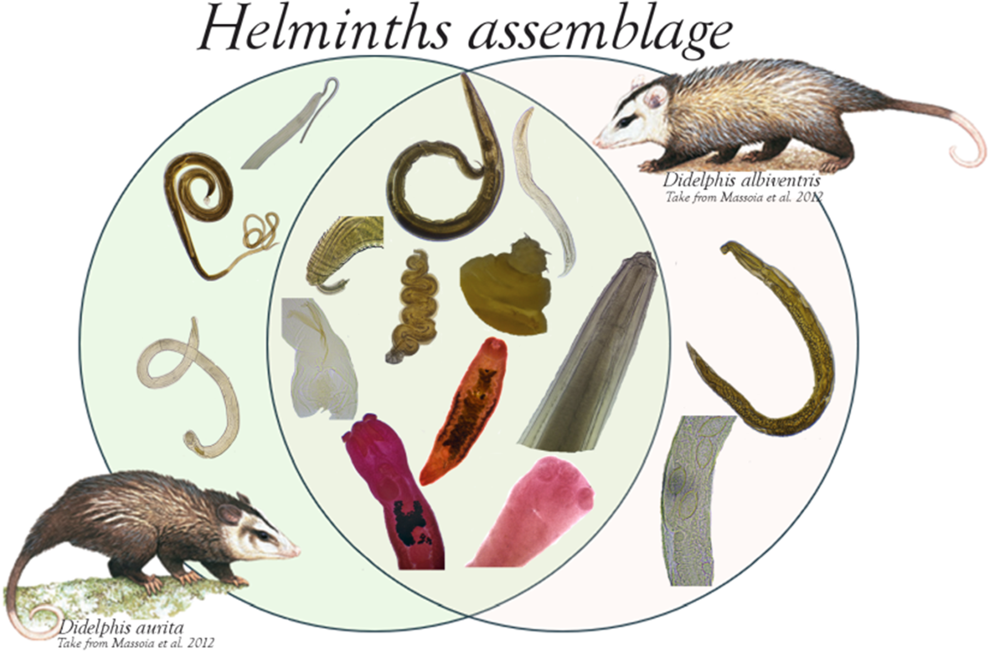
In Misiones, opossums comprise 13% of the wild mammalian diversity and they are represented by 9 genera and 15 species: Caluromys Allen, 1900, Chironectes Illiger, 1811, Didelphis Linnaeus, 1758, Gracilinanus Gardner y Creighton, 1989, Lutreolina Thomas, 1910, Metachirus Burmeister,1854, Micoureus Lesson, 1842, Monodelphis Burnett, 1830, and Philander Tiedemann, 1808 (Massoia et al., Reference Massoia, Chebez and Bosso2012). The white-eared opossum (Didelphis albiventris Lund, 1840) and the southern black-eared opossum D. aurita Wied-Neuwied, 1826 are the so called medium sized opossums, frequently found in human dwellings and peripheral areas in north Misiones (Massoia et al., Reference Massoia, Chebez and Bosso2012).
Didelphis albiventris is distributed in Brazil, Bolivia, Paraguay, Uruguay and Argentina (Wilson and Reeder, Reference Wilson and Reeder2005). In Argentina, this species is distributed from north to the central part of the country (Chemisquy and Gabriel, Reference Chemisquy and Martin2019) with an expanding presence toward the south (Pastrán-López et al., Reference Pastrán-López, Rivero-Castro, Ruiz-Estebez, Amoni-Sacchi and Sánchez-Castro2022). This opossum is found in diverse habitats like forests and grasslands often associated with human-altered landscapes (Massoia et al., Reference Massoia, Chebez and Bosso2012). Didelphis aurita is distributed along the coast of Brazil, from Bahia to Rio Grande do Sul, east of the lower Paraguay river and the Argentinian Atlantic Forest (AAF) (Wilson and Reeder, Reference Wilson and Reeder2005). Unlike D. albiventris, it is associated in well-preserved native forests, both in continuous areas and remnants (Massoia et al., Reference Massoia, Chebez and Bosso2012; Chemisquy and Gabriel, Reference Chemisquy and Martin2019). Both species are omnivorous and have opportunistic feeding behaviour, with a diet consisting of small vertebrates, invertebrates, seeds and fruits (Massoia et al., Reference Massoia, Chebez and Bosso2012). Additionally, these opossums may consume garbage remnants of human consumption, as well as food available inside the forest (Bezerra-Santos et al., Reference Bezerra-Santos, Ramos, Campos, Dantas-Torres and Otranto2021).
In Argentina, studies on helminths in opossums are scarce and sporadic over time (Hartmann, Reference Hartmann2023). At present, about 27 helminth species are known to be present in the most common species of opossums, mostly from central Argentina (Santa Cruz et al., Reference Santa Cruz, Borda, Montenegro, Gómez, Prieto and Schebler1999; Lunaschi and Drago, Reference Lunaschi and Drago2007; Castaño Zubieta et al., Reference Castaño Zubieta, Ruiz, Morici, Lovera, Fernández, Caracostantogolo and Cavia2014; Montes de Oca et al., Reference Montes de Oca, Dominguez, Lammel and Cavia2024) and a few records from a rare species in the Northwest Argentina (Navone, Reference Navone1989; Navone et al., Reference Navone, Suriano and Pujol1991; Navone and Suriano, Reference Navone and Suriano1992). To our knowledge, in the AF there are several studies that report helminths in D. aurita and D. albiventris (see Antunes, Reference Antunes2005; Ramos et al., Reference Ramos, Santos, Freitas, Correa, Kempe, Morgado, Aguiar, Wolf, Rossi, Sinkoc and Pacheco2016; Costa-Neto et al., Reference Costa-Neto, Cardoso, Boullosa, Maldonado and Gentile2018; Bezerra-Santos et al., Reference Bezerra-Santos, Ramos, Campos, Dantas-Torres and Otranto2021). However, in Misiones there is only one record of Martínez (Reference Martínez1986) mentioning the trematode Duboisella proloba Baer, 1938 in D. albiventris.
As opossums are frequent in urban and rural environments, live in close relationship with humans and domestic animals, they are considered potential reservoirs of many infectious agents (i.e. Trypanossoma cruzi, Leishmania infantum, Ancylostoma caninum, Angiostrongylus cantonensis, among others) (Bezerra-Santos et al., Reference Bezerra-Santos, Ramos, Campos, Dantas-Torres and Otranto2021). Several studies have shown that opossums are involved in the transmission of parasites of animal health concern, playing an underestimated role in the epidemiology of parasitic diseases affecting domestic animals (Bezerra-Santos et al., Reference Bezerra-Santos, Ramos, Campos, Dantas-Torres and Otranto2021). In this study we proposed to work toward filling this knowledge gap providing new and updated records of helminth species from D. albiventris and D. aurita in the AAF.
Materials and methods
Study site
Our study was conducted between September 2021 and June 2023 in the Department of Puerto Iguazu in the north of Misiones province. Sampling was conducted at various sites, including the routes through Iguazu National Park (National Routes (NR) 12 and 101), Colonia Wanda (NR 12 and Provincial Route (PR) 19), and Puerto Esperanza (NR 12), as well as the urban and rural areas surrounding these cities (Figure 1).

Figure 1. Map of the Iguazú Department. In red is marked the route of National Routes 12 and 101 and Provincial Route 19. Individuals of Didephis albiventris collected are marked with green dots and those of D. aurita with purple dots.
Sample collection
We collected opportunistically opossum carcasses recently dead, including those from roadkill, dog attacks, or other causes. Specimens were collected in plastic bags and necropsied in the laboratory. The body cavity and viscera were thoroughly examined using a magnifying glass to facilitate observation. We collected and preserved all parasites found. Nematodes and acanthocephalans were fixed in 10% formalin and preserved in 70% ethanol. For morphological studies they were cleared in Amman’s lactophenol and studied with a Leica DM500 microscope equipped with a drawing tube. Trematodes and cestodes were placed between 2 slides with 2% formalin until they were flattened; and preserved in 70% ethanol. For morphological studies specimens to be studied were stained with 1:6 dilutions in 96% ethanol of hydrochloric carmine, dehydrated in a series of alcohol ranging 70%–85%–95%–100% ethanol, rinsed in eugenol, and mounted in Canada balsam. Measurements are given in millimetres (mm) unless other units are stated. Photographs were taken with a Zeiss Primo Star microscope with a compact digital camera.
Voucher specimens were deposited in the Colección de Helmintos Museo de La Plata, Argentina (CHMLP-he) (see Appendix), and IBS authors collection. Host acronyms correspond to the field numbers BH (Barbara Hartmann) and M (Dante Di Nucci), and to the Colección de Mastozoología del Laboratorio de Genética Evolutiva – Dr Claudio Bidau (CM-LGE) from the Instituto de Biología Subtropical IBS (see Appendix).
Populations parameters
We analysed parameters at the component population level. Quantitative parameters of prevalence (P), mean abundance (MA) and mean intensity (MI) were calculated following Bush et al. (Reference Bush, Lafferty, Lotz and Shostak1997). For the prevalence, a 95% confidence interval (CI) was estimated using Sterne’s method; a bias-corrected and accelerated 95% bootstrap CI (Bca) with 2000 replicates was applied for calculation of MI and MA, according to Reiczigel et al. (Reference Reiczigel, Marozzi, Fábián and Rózsa2019). The Quantitative Parasitology web software was used to calculate these descriptors (Reiczigel et al., Reference Reiczigel, Marozzi, Fábián and Rózsa2019).
Results
Thirteen specimens of D. albiventris and 11 of D. aurita were collected. Three species of Platyhelminthes, 10 species of nematodes and 1 species of Acanthocephala were identified. In Table 1 we detail the helminth species and the parameters (P, MI and MA) recovered from both, D. albiventris and D. aurita in the present study. Below is listed the helminths species including a brief description with their measurements.
Table 1. Population parameters of the helminths parasitizing Didelphis albiventris and D. aurita from Northern Misiones

* †Helminths with heteroxenous life cycle. SI: Small intestine; LI: Large intestine; C: Caecum; S: Stomach; T: Trachea; S: Stomach.
* Only one infected host in the sample, thus CI cannot be calculated.
Taxonomic aspects
Phylum Platyhelminthes Minot, 1876
Class Trematoda Rudolphi. 1808
Superfamily Brachylaimoidea Joyeux and Foley, 1930
Family Brachylaimidae Joveux and Foley, 1930
Brachylaima migrans Dujardin, 1845 (Fig. 2A)

Figure 2. Photographs of helminths: A. Brachylaima migrans. B. Anterior end of Rhopalias coronatus. C. Trichuris minuta: 1. Posterior end. 2. Detail of the spicular sheath. D. Mathevotaenia sp: 1. Scolex. 2. Immature proglotids. 3. Mature gravid proglotids. E. Detail of spicular sheath of Trichuris didelphis. F. Capillaria sp. 1: 1. Posterior end. 2. Detail of spicular sheath and spicule. G. Detail of the vulva and eggs of Capillaria sp. 2. H. Globocephalus marsupialis: 1. Detail of the anterior region. 2. Tail, detail of bursa and spicule. I. Tail of Viannaia viannai. J. Travassostrongylus orloffi: 1. Male tail, detail of spicules, gubernaculum, and telamon. 2. Female, detail of the vulva. K. Aspidodera raillieti: 1. Anterior region. 2. Tail, detail of spicules, gubernaculum, and sucker. L. Travassostrongylus callis: 1. Male tail, detail of spicules, gubernaculum, and telamon. 2. Female vulva with cuticular cap. M. Cruzia tentaculata, male tail, detail of spicules and gubernaculum. N. Oligacanthorhynchus microcephala proboscis.
Description (n = 5): Trematode small, whitish, elongated subcylindrical body 3.15–4.31 long by 0.60–0.90 wide. Oral and ventral suckers globular, nearly equal, 0.40–0.50 by 0.30–0.60 and 0.30–0.45 by 0.30–0.43, respectively. Oesophagus 0.20–0.30 long by 0.15–0.20 wide. Testes in tandem, located in the posterior third of the body, anterior testis 0.20–0.40 long by 0.20–0.35 wide, posterior testes 0.25–0.45 long by 0.20–0.30 wide. Ovary intertesticular, 0.15–0.25 long by 0.20–0.30 wide, the oviduct 0.2–0.6 long is continued by the uterus 1.75–2.5 long, which open into the genital pore. Eggs elliptical, with one side slightly flattened and the other convex, 0.02 long by 0.01 wide.
Taxonomic summary
Host: Didelphis albiventris (BH01, BH06, BH10, M177/22, M190/22, M211/23), Didelphis aurita (BH09, BH20, BH24, BH26, BH32, M220/23).
Infection site: Small intestine
Localities: Puerto Esperanza, Colonia Wanda, and Puerto Iguazú, Misiones, Argentina (see appendix).
Deposited material: MLP-He 8141 (1 specimen in a slide).
Remarks: The morphological characters are consistent with the description given by Boero and Boehringer (Reference Boero and Boehringer1967). Brachylaima migrans has been recorded across Argentina as a parasite of D. albiventris and Lutreolina crassicaudata Desmarest, 1804 (see Boero and Boehringer, Reference Boero and Boehringer1967; Martínez, Reference Martínez1986; Santa Cruz, Reference Santa Cruz2006; Lunaschi and Drago, Reference Lunaschi and Drago2007). In Brazil, this species was reported for D. albiventris (see Silva and Costa, Reference Silva and Costa1999; Antunes, Reference Antunes2005) and D. aurita (see Costa-Neto et al., Reference Costa-Neto, Cardoso, Boullosa, Maldonado and Gentile2018; Gentile et al., Reference Gentile, Costa-Neto, Cardoso and Maldonado2022). This is the first record for D. aurita in Argentina.
Superfamily Echinostomatoidea Looss, 1899
Family Rhopaliidae Looss, 1899
Rhopalias coronatus (Rudolphi, 1819) Stiles and Hassall, 1898 (Fig. 2B)
Description (n = 5): Trematodes with elongated, spiny body of 7.35–10.85 long by 1.20–1.50 wide. Specimens possess a pair of armed proboscides with spines, which can be invaginated into a muscular pouch. The pouches open to the exterior on each side of the oral sucker. Subterminal oral sucker. Acetabulum larger than the oral sucker, at 1.40–2.75 from the anterior end. Testes oval, in tandem located in the posterior half of the body, anterior testis 0.50–0.85 long by 0.30–0.60 wide and posterior testes 0.6–0.9 long by 0.3–0.4 wide. Ovary oval, pretesticular, 0.45–0.95 long by 0.4–0.85 wide. Genital pore median, pre-acetabular. Vitelline follicles in lateral fields in posterior third of the body. Eggs oval to elliptical, 0.09–0.1 long by 0.05–0.06.
Taxonomic summary
Host: Didelphis albiventris (M190/22, M211/23), Didelphis aurita (BH07, BH08, BH20).
Infection site: Small intestine
Locality: Puerto Iguazú, Misiones, Argentina.
Deposited material: MLP-He 8142 (1 specimen on a slide).
Remarks: Morphology of our specimens is consistent with the description given by Haverkost and Gardner (Reference Haverkost and Gardner2008) and Chero et al. (Reference Chero, Sáez, Mendoza-Vidaurre, Iannacone and Cruces2017). However, they are larger worms, the tentacle sacs extend far beyond the posterior margin of the pharynx, and the tentacle spines and oral spines are difficult to observe compared to those reported by Haverkost and Gardner (Reference Haverkost and Gardner2008). Species of Rhopalias are parasites of the small intestine of marsupials from the Nearctic and Neotropical region (Haverkost and Gardner, Reference Haverkost and Gardner2008). Rhopalias coronatus has been reported in D. albiventris and L. crassicaudata from Argentina (see Boero and Boehringer, Reference Boero and Boehringer1967; Lombardero and Moriena, Reference Lombardero and Moriena1973; Martínez et al., Reference Martínez, Brandetti and Boero1973; Martínez, Reference Martínez1986; Haverkost and Gardner, Reference Haverkost and Gardner2008; Jiménez et al., Reference Jiménez, Campbell, Byles, Scheibel and Gardner2024). It was also reported from D. albiventris from Brazil and Paraguay (see Haverkost and Gardner, Reference Haverkost and Gardner2008; Marinho de Quadros et al., Reference Marinho de Quadros, Müller, Barbosa de Moura, Rodríguez, Martins, Filippi and Mazzoni2016; Zabott et al., Reference Zabott, Pinto, Viott, Gruchouskei and de Barros Bittencourt2017; Teodoro et al., Reference Teodoro, Cutolo, Motoie, da Silva Meira-strejevitch, Pereira-Chioccola, Fernandes Mendes and Marques Allegretti2019; Jiménez et al., Reference Jiménez, Campbell, Byles, Scheibel and Gardner2024); D. aurita (see Costa-Neto et al., Reference Costa-Neto, Cardoso, Boullosa, Maldonado and Gentile2018; Gentile et al., Reference Gentile, Costa-Neto, Cardoso and Maldonado2022) from Brazil, and Philander opossum Linnaeus, 1758 from Bolivia (Haverkost and Gardner, Reference Haverkost and Gardner2008; Jiménez et al., Reference Jiménez, Campbell, Byles, Scheibel and Gardner2024). This is the first record for D. aurita in Argentina.
Class Cestoda Rudolphi 1808
Orden Cyclophyllidea van Beneden in Braun, 1900
Family Anoplocephalidae Blanchard, 1891
Mathevotaenia sp. (Fig. 2D1-D3)
Description (n = 5): Small cestodes, 4.95–5.60 in total length, consisting of 19–25 proglottids. Maximum width 0.35–0.45 attained in gravid segments. Scolex unarmed, poorly demarcated from strobila, 0.25–0.30 long by 0.40–0.50 wide. Oval suckers, with thin muscular walls, 0.15–0.2 long by 0.15–0.2 wide. In the specimens studied herein, segmentation begins immediately after scolex. Proglottids trapezoidal with laterally expanded posterior edges. Mature eggs concentrated along the lateral margin of the proglottids. Egg capsule 0.2 by 0.2.
Taxonomic summary
Hot: D. albiventris (M211/23) and D. aurita (BH20).
Infection site: Small intestine.
Locality: Puerto Iguazú, Misiones, Argentina.
Deposited material: Specimens under study.
Remarks: The characteristics of these cestodes correspond to those mentioned by Campbell et al. (Reference Campbell, Gardner and Navone2003) for Mathevotaenia spp.: small cestodes, craspedote proglottids, scolex unarmed. However, the worms studied herein are smaller than those mentioned by Campbell et al. (Reference Campbell, Gardner and Navone2003). Mathevotaenia argentinensis (Campbell et al., Reference Campbell, Gardner and Navone2003) and M. sanmartini Jimenez, Cambell & Gardner, 2008 present 135–163 proglottids and more than 200, respectively, while M. bivittata (Janicki, 1904) has 37–49 proglottids. Our specimens have fewer proglottids, and the position of the ovary and testes differs from M. bivittata, meaning that these specimens could be a new species. More specimens should be studied to confirm this.
In Argentina, M. bivittata (Campbell et al., Reference Campbell, Gardner and Navone2003) and M. sanmartini were recorded for Marmosa cinerea and M. argentinensis was recorded for Thylamys pallidior (see Jiménez et al., Reference Jiménez, Braun, Campbell and Gardner2008). In Brazil, M. bivittata was reported parasitizing D. albiventris (see Justo et al., Reference Justo, Fernandes, Knoff, Cárdenas and Cohen2017). Other species of the genus Didelphis have been mentioned as hosts for Mathevotaenia sp. in Mexico and French Guiana (Monet-Mendoza et al., Reference Monet-Mendoza, Osorio-Sarabia and Garcia-Prieto2005; Jiménez et al., Reference Jiménez, Catzeflis and Gardner2011). This is the first report of Mathevotaenia sp. from D. aurita.
Phylum Nemata Rudolphi, 1808
Order Trichinellida Hall, 1916
Family Trichuridae Ramson, 1911
Trichuris minuta (Rudolphi, 1819) (Babero, Reference Babero1960) (Fig. 2C1, C2)
Description (6 males and 4 females): Mouth without lips; stichosome with 1 row of stichocytes. Bacillary band present. Spicule 0.82–1.14 long, spicule sheath 0.11–0.19 long, with spines less than 1 μm long, spicule tube 0.53–1.14 long, ejaculatory duct 0.95–1.65 long and testes 3.60–5.95 long. Vulva located at the 9.70–14.41 from the anterior end, vagina 0.90–1.60 long, ovary located at the 1.02–1.20 from the posterior end, uterus monodelphic, terminal anus. Eggs 0.06–0.07 long by 0.03–0.04 wide.
Taxonomy summary
Host: Didelphis albiventris (BH05, BH06, BH10, BH35, M174/22, M177/22, M211/23), Didelphis aurita (BH07, BH08, BH09, BH19, BH20, BH24, BH32).
Infection site: Large intestine and caecum.
Localities: Puerto Esperanza, Colonia Wanda, and Puerto Iguazú; Misiones, Argentina.
Deposited material: MLP-He 8143 (5 males and 5 females).
Remarks: Our specimens are morphologically like those described by Babero (Reference Babero1960). Trichuris minuta was originally described by Rudolphi (1819) parasitizing Caluromys phylander [originally mentioned as Didelphis cayopollin (Schreber, 1777)] in Brasilia, Brazil (Rudolphi, 1819). Later, Babero re-described the species from D. virginiana from Georgia, USA. More recently, this species was recorded in Brazil as a parasite of D. albiventris (Antunes, Reference Antunes2005) and D. aurita (see Noronha et al., Reference Noronha, Vicente and Pinto2002; Costa-Neto et al., Reference Costa-Neto, Cardoso, Boullosa, Maldonado and Gentile2018). This is the first report of T. minuta for Argentina.
Trichuris didelphis (Babero, Reference Babero1960) (Fig. 2E)
Description (4 males): Anterior end very thin, mouth without lips. Stichosoma occupies approximately two-thirds of the length of the body; the body widens at the level of esophago-intestine junction. Spicule 0.89–0.20 long. Spicule sheath extending 0.20–0.30 beyond rear end of body, globular, pear-shaped in outline, with small spines less than 1 µm long. Cloaca 0.25–0.43 long, thick and muscular. Spicule tube 0.15–0.20 long. Ejaculatory duct 1.50–1.80 long and vas deferens 2.40–3.8 long.
Taxonomic summary
Host: Didelphis aurita (BH26, BH32, M220/23).
Infection site: Caecum.
Locality: Puerto Iguazú, Misiones, Argentina.
Deposited material: Specimens under study.
Remarks: This species was originally described by Babero (Reference Babero1960) from D. virginiana in Georgia, USA. In Brazil, it was also recorded in D. albiventris (see Silva and Costa, Reference Silva and Costa1999; Antunes, Reference Antunes2005) and D. aurita (see Costa-Neto et al., Reference Costa-Neto, Cardoso, Boullosa, Maldonado and Gentile2018; Gentile et al., Reference Gentile, Costa-Neto, Cardoso and Maldonado2022). While in Argentina, it was recorded in D. albiventris (see Santa Cruz, Reference Santa Cruz2006). Lombardero and Moriena (Reference Lombardero and Moriena1973) reported the presence of Trichuris sp. in D. albiventris from Corrientes province. The drawings provided by these authors closely resemble T. didelphis, indicating that it might be the same species. This species was also reported in Mexico (Acosta-Virgen et al., Reference Acosta-Virgen, López-caballero, García-Prieto and Mata-López2015). This is the first report of T. didelphis parasitizing D. aurita in Misiones.
Family Capillaridae Raillet, 1915
Capillaria sp. 1 (Fig. 2F1, F2)
Description (5 females and 2 males): Small, slender-bodied nematodes, 11.2–16.50 total length; esophagus 5–5.3 long by 0.03–0.06 wide with well differentiated sticocytes and visible bacillary band; spicule 0.97–1 long and 0.01 wide, surrounded by a 0.67–0.9 spicule tube, covered by small cuticular spines; vulva located 4.45 from anterior region; eggs elongated barrel-shaped, colourless shell and operculum slightly projected outward, 0.05 by 0.03.
Taxonomic summary
Host: Didelphis aurita (BH08, BH32)
Infection site: Trachea, esophagus, stomach and small intestine.
Locality: Puerto Iguazú, Misiones, Argentina.
Deposited material: Specimens under study (retained by authors).
Remarks: Based on the morphological characters and the site of infection, we identified the specimen belonging to the genus Capillaria. In Brazil, several authors reported the presence of Capillaria sp. in the digestive tract of D. albiventris and other species of Didelphidae (Vicente et al., Reference Vicente, Oliveira Rodrigues, Correa Gomes and Magalhaes Pinto1997; Silva and Costa, Reference Silva and Costa1999; Noronha et al., Reference Noronha, Vicente and Pinto2002). Once again, these authors did not include good descriptions of the specimens. The genus Capillaria has a great diversity of species and many of them have been recorded from wild mammals (Butterworth and Beverley-Burton, Reference Butterworth and Beverley-Burton1977). Species identification of Capillaria is challenging due to the scarcity of males, the indistinct morphological traits of females and, in many cases, the limited availability of descriptions (López-Neyra, Reference López-Neyra1946). Moreover, Moravec (Reference Moravec1982) argues that many of the descriptions of the genus Capillaria are erroneous. Herein, our specimens clearly correspond to Capillaria sp. because they were found in the digestive tract (Moravec, Reference Moravec1982). In Didelphidae previous studies have reported the presence of C. longicauda Freitas and Lent, 1935 in P. opossum and D. marsupialis (see López-Neyra, Reference López-Neyra1946; Jiménez et al., Reference Jiménez, Catzeflis and Gardner2011). The spicule of C. longicauda has been described as poorly chitinized, transparent, and difficult to observe (López-Neyra, Reference López-Neyra1946). However, in this study the spicule was highly visible and well-chitinized, similarly to Capillaria eberthi. Further material and a revision of the specimens should be necessary to elucidate the identity of the species.
Capillaria sp. 2 (Fig. 2G)
Description (4 females): Nematodes small and slender; total length 7.75–10.05; oesophagus 3.15–4.25 long and 0.02–0.03 wide, with well-differentiated sticocytes, bacillary band absent; vulva at 3.85–4.3 from anterior end; eggs rounded, barrel-shaped, colourless shell with slightly outward projecting opercula, 0.06–0.07 by 0.03–0.04.
Taxonomy summary
Host: Didelphis albiventris (M177/22; M190/22)
Infection site: Stomach and small intestine.
Localities: Puerto Iguazú, Misiones, Argentina.
Deposited material: Specimens under study (retained by authors).
Remarks: Based on the morphological characters and the site of infection, we identified the specimen belonging to the genus Capillaria. In this case, only female specimens were recovered. Capillaria sp. 2 differ from Capillaria sp. 1 in body size, as they are considerably smaller; by the absence of the bacillary band, which was clearly observed in Capillaria sp. 1, and by the shape and size of the eggs, which are more rounded and larger than those of Capillaria sp. 1.
Order Strongylida Raillet y Henry, 1913
Family Ancylostomatidae Looss, 1905
Globocephalus marsupialis (Freitas de Teixeira and Lent, Reference Freitas de Teixeira and Lent1936) (Fig. 2H1, H2)
Description (10 males and 10 females): White body with transversely striated cuticle. Subglobular stoma, with a pair of large sub-ventral teeth near the base of the capsule. Well-developed claviform muscular oesophagus. Spicules 0.32–0.45 long and gubernaculum 0.03–0.07 long. Female with amphidelphic uterus. Vulva at 4–5.25 from the anterior end. Eggs 0.084–0.11 long by 0.05–0.06 wide.
Taxonomy summary
Host: Didelphis albiventris (M190/22)
Infection site: Small intestine.
Locality: Puerto Iguazú, Misiones, Argentina.
Deposited material: MLP-He 8145 (5 males and 5 females).
Remarks: The specimens herein studied are consistent with the morphological characteristics given by Freitas de Teixeira and Lent (Reference Freitas de Teixeira and Lent1936) parasitizing Metachirus myosurus Temminck, 1824 from Brazil. It was also recorded in D. aurita (see Costa-Neto et al., Reference Costa-Neto, Cardoso, Boullosa, Maldonado and Gentile2018; Gentile et al., Reference Gentile, Costa-Neto, Cardoso and Maldonado2022). This is the first report of G. marsupialis parasitizing D. albiventris in Argentina.
Family Viannaiidae Neveu-Lemaire, 1944
Viannaia viannai (Travassos, Reference Travassos1914) (Fig. 2I)
Description (8 males and 6 females): nematodes small and thin, body curled around its own axis. Synlophe without lateral or dorsal ridges. Cephalic extremity with a highly developed cuticular expansion. Males with highly developed caudal bursa, with 2 slightly asymmetrical lateral lobes and highly developed dorsal lobe. Bursa 2–1–2 type. Spicules 0.12–0.158 long. Females monodelphic. Vulva at 1.25–2.8 from posterior end. Eggs 0.05–0.07 long by 0.04–0.05 wide.
Taxonomic summary
Host: Didelphis albiventris (BH35; M190/22, M211/23), Didelphis aurita (BH07, BH08, BH09, BH21, BH24, BH32, BH34, M220/23).
Infection site: Small intestine.
Localities: Puerto Iguazú, Misiones, Argentina.
Deposited material: MLP-He 8144 (5 males and 5 females).
Remarks: The specimens fit the description provided by Travassos (Reference Travassos1914). In Brazil, V. viannai was reported in D. aurita (see Travassos, Reference Travassos1914) and P. opossum (see Corrêa Gomes et al., Reference Corrêa Gomes, da Cruz R, Vicente and Magalhães Pinto2003); and in Bolivia it was mentioned parasitizing 6 species of Didelphidae (see Jiménez et al., Reference Jiménez, Campbell, Byles, Scheibel and Gardner2024). It was also reported from Peru, Mexico, Venezuela, and French Guyana (Guerrero, Reference Guerrero1985; Monet-Mendoza et al., Reference Monet-Mendoza, Osorio-Sarabia and Garcia-Prieto2005; Jiménez et al., Reference Jiménez, Catzeflis and Gardner2011). This is the first report of V. viannai parasitizing Didelphis spp. in Argentina.
Travassostrongylus orloffi Travassos, 1935 (Fig. 2J1, J2).
Description (4 males and 5 females): Small nematodes. Cuticle with transverse striations and longitudinal lines; synlophe with 10 symmetric crests oriented from right to left. Cephalic cuticular dilatation of 0.09–0.13. Bursa 2–2–1 type. Spicules subequal, complex, 0.12–0.14 long by 0.02–0.04 wide, distal end bifid; gubernaculum 0.06–0.11 long; conical telamon 0.03–0.07 long. Vulva 1.05–1.2 from posterior end, anus 0.1–0.14 from posterior end. Eggs 0.05–0.06 long by 0.04–0.05 wide.
Taxonomic summary
Host: Didelphis aurita (BH32)
Infection site: Small intestine
Localities: Puerto Iguazú, Misiones, Argentina.
Deposited material: Specimens under study (retained by authors).
Remarks: Our specimens are morphologically similar to those described by Diaw (Reference Diaw1976). Descriptions for this species by Travassos (Reference Travassos1937) and Diaw (Reference Diaw1976) mention the presence of a subconical telamon. In this work, we also note that the telamon exhibits bilateral projections at one-third of its height. In Brazil, T. orloffi was reported for D. albiventris (see Silva and Costa, Reference Silva and Costa1999; Antunes, Reference Antunes2005) and D. aurita (see Costa-Neto et al., Reference Costa-Neto, Cardoso, Boullosa, Maldonado and Gentile2018; Gentile et al., Reference Gentile, Costa-Neto, Cardoso and Maldonado2022). It was also found in French Guyana (Diaw, Reference Diaw1976) and Mexico (Scheibel et al., Reference Scheibel, Catzeflis and Jimenez2014). This is the first record of Travassostrongylus orloffi for D. aurita in Argentina.
Travassostrongylus callis (Travassos, Reference Travassos1914) Orloff, 1933 (Fig. 2L1, L2)
Description: (7 males and 5 females): Nematodes small, cuticle with transverse and longitudinal striations, cephalic cuticular dilatation 0.1–0.11. Bursa 2–2–1 type. Spicules subequal, complex, 0.1–0.5 long and 0.01–0.02 wide, gubernaculum 0.04–0.05 long, conical telamon 0.03–0.05 long, with a left hook. Vulva located 1–1.4 from hind end, with a cuticular cap. Anus 0.1–0.13 from posterior end. Eggs 0.05 × 0.03.
Taxonomic summary
Host: Didelphis albiventris (M211/23), Didelphis aurita (BH32, M220/23)
Infection site: Small intestine
Localities: Puerto Iguazú, Misiones, Argentina.
Deposited material: Specimens under study (retained by authors).
Remarks: Our specimens are similar with those described by Travassos (Reference Travassos1937) and Diaw (Reference Diaw1976). Travassostrongylus callis was originally found in D. aurita from Brazil (Travassos, Reference Travassos1914). Later, it was mentioned from other localities from Brazil (Noronha et al., Reference Noronha, Vicente and Pinto2002), and recently Jiménez et al. (Reference Jiménez, Campbell, Byles, Scheibel and Gardner2024) mentioned it for Chironectes minimus Zimmermann, 1780 in Bolivia. It was also found parasitizing D. marsupialis in French Guiana (Diaw, Reference Diaw1976), and Panama (Scheibel et al., Reference Scheibel, Catzeflis and Jimenez2014). This is the first report in D. albiventris and D. aurita from Argentina.
Order Ascaridida Chabaud, 1965
Superfamily Heterakoidea Railliet and Henry, 1912
Family Aspidoderidae Skrajabin y Schikhobalova, 1947
Aspidodera raillieti Travassos, 1913 (Fig. 2K1, K2)
Description (9 males and 9 females): Anterior end with a cap with 6 longitudinal loops or cords, 3 of them on the interlips and 3 on each lip. Oesophagus with a terminal bulb. Spicules similar in shape and size, 0.55–0.93 long. Gubernaculum 0.15–0.22 long. Cloaca at 0.28–0.38 from posterior end. Vulva at 1.90–2.65 from posterior end, amphidelphic uterus. Eggs 0.05–0.07 long by 0.04–0.05 wide.
Taxonomic summary
Host: Didelphis albiventris (BH35, M174/22), Didelphis aurita (BH07, BH08, BH09, BH20, BH21, BH32, M220/23).
Infection site: Large intestine and caecum
Localities: Puerto Iguazú, Misiones, Argentina.
Deposited material: MLP-He 8147 (5 males and 5 females).
Remarks: Nematodes similar in morphology to those described by Portes Santos et al. (Reference Portes Santos, Lent and Correa Gomes1990) and (Chagas-Moutinho et al., Reference Chagas-Moutinho, Oliveira-Menezes, Cárdenas and Lanfredi2007). The specimens characterized by Chagas-Moutinho et al. (Reference Chagas-Moutinho, Oliveira-Menezes, Cárdenas and Lanfredi2007) are relatively smaller and we consider that the differences are induced by the host and geographic distance. Aspidodera raillieti was also recorded in several species of marsupials from Brazil (Costa-Neto et al., Reference Costa-Neto, Cardoso, Boullosa, Maldonado and Gentile2018; Gentile et al., Reference Gentile, Costa-Neto, Cardoso and Maldonado2022), Bolivia, Paraguay and Mexico (Jiménez et al., Reference Jiménez, Campbell, Byles, Scheibel and Gardner2024), French Guiana (Jiménez et al., Reference Jiménez, Catzeflis and Gardner2011), Peru (Polo-González et al., Reference Polo-González, Sánches and Pacheco2019), and other localities. In Argentina, A. raillieti was only recorded in D. albiventris (see Lombardero and Moriena, Reference Lombardero and Moriena1973; Navone and Suriano, Reference Navone and Suriano1992). This is the first record for D. aurita in Argentina.
Family Kathlaniidae Lane, 1914
Cruzia tentaculata (Rudolphi, 1819) (Travassos, Reference Travassos1922) (Fig. 2M)
Description (20 males and 20 females): Oral opening surrounded by 3 lips, 1 dorsal and 2 latero-ventral; inner margin of lips with small, serrated structures. Oesophagus ending in a bulb with an intestinal caecum projecting anteriad beyond the bulb. Spicules 0.80–1.20 long. Gubernaculum 0.18–0.40. Cloaca at 0.10–0.20 from posterior end. Vulva located at the middle of the body 4.35–9.40 from the anterior end. Anus at 0.55–1.90 from the posterior end. Eggs 0.10–0.15 long by 0.5–0.07 wide.
Taxonomic summary
Host: Didelphis albiventris (BH01, BH05, BH06, BH10, M128/22, BH35, M82/21, M129/22, M174/22, M177/22, M190/22, M211/23), Didelphis aurita (BH07, BH08, BH09, BH19, BH20, BH21, BH24, BH26, BH32, BH34, M220/23).
Infection site: Large intestine and caecum.
Localities: Puerto Esperanza, Colonia Wanda, Puerto Iguazú, Misiones, Argentina.
Deposited material: MLP-He 8146 (5 males and 5 females).
Remarks: Cruzia tentaculata was originally described parasitizing D. aurita from Brazil (Travassos, Reference Travassos1922). After that, it was recorded in several South American didelphids (see Noronha et al., Reference Noronha, Vicente and Pinto2002; Corrêa Gomes et al., Reference Corrêa Gomes, da Cruz R, Vicente and Magalhães Pinto2003; Adnet et al., Reference Adnet, Anjos, Menezes-Oliveira and Lanfredi2009; Costa-Neto et al., Reference Costa-Neto, Cardoso, Boullosa, Maldonado and Gentile2018; Jiménez et al., Reference Jiménez, Campbell, Byles, Scheibel and Gardner2024). In Argentina, it is mentioned parasitizing D. albiventris and L. crassicaudata (see Boero and Boehringer, Reference Boero and Boehringer1967; Santa Cruz, Reference Santa Cruz2006). This is the first record for D. aurita in Argentina.
Order Spirurida Railliet, 1915
Family Physalopteridae Railliet, 1893
Turgida turgida (Rudolphi 1819) Travassos, 1919
Description (5 males and 5 females): Large nematodes with a rigid whitish body, covered by a thick cuticle with fine transverse striations. Oral opening surrounded laterally by 2 symmetrical, semi-domed lips or pseudolabia, composed of 3 fused lips that are flattened on the inner part. A cephalic collarette, formed by a deep fold surrounds the pseudolabia. Male tail with caudal alae with 22 papillae: 4 pairs of pedunculated papillae are placed on the caudal alae; 3 papillae are located anteriorly to the cloaca; spicules 0.35–0.60 long; tail 0.3–0.9 length. Vulva is located below the end of the oesophagus at 4.84–9.10 from the anterior end; tail 0.3–0.7 length.
Taxonomic summary
Host: Didelphis albiventris (BH01, M129/22, M190/22, M211/23), Didelphis aurita (BH07, BH19, BH20, BH21, BH26).
Infection site: Stomach
Localities: Colonia Wanda, and Puerto Iguazú, Misiones, Argentina.
Deposited material: MLP-He 8148 (5 males and 5 females)
Remarks: Turgida turgida was recorded parasitizing Didelphis spp. from North and South America (Humberg et al., Reference Humberg, Tavares, Paiva, Oshiro, Bonamigo, Júnior and Oliveira2011). Our specimens are morphologically like those described by Travassos (Reference Travassos1920), Matey et al. (Reference Matey, Kuperman and Kinsella2001), and Humberg et al. (Reference Humberg, Tavares, Paiva, Oshiro, Bonamigo, Júnior and Oliveira2011). In Brazil, T. turgida was recorded in D. albiventris, D. aurita, C. minimus and P. quica Temminck, 1824 (see Noronha et al., Reference Noronha, Vicente and Pinto2002; Humberg et al., Reference Humberg, Tavares, Paiva, Oshiro, Bonamigo, Júnior and Oliveira2011; Costa-Neto et al., Reference Costa-Neto, Cardoso, Boullosa, Maldonado and Gentile2018); from Bolivia in D. albiventris and P. opossum (see Jiménez et al., Reference Jiménez, Campbell, Byles, Scheibel and Gardner2024), while in Argentina it was reported parasitizing D. albiventris and L. crassicaudata (see Boero and Boehringer, Reference Boero and Boehringer1967; Navone and Suriano, Reference Navone and Suriano1992; Santa Cruz, Reference Santa Cruz2006). This is the first record for D. aurita in Argentina.
Phylum Acanthocephala Kohlreuther, 1771
Clase Archiacanthocephala Meyer, 1931
Orden Oligacanthorhynchida Petrochenko, 1956
Familia Oligacanthorhynchidae Southwell et Macfie, 1925
Oligacanthorhynchus microcephalus (Rudolphi, 1819) Schmidt, 1972 (Fig. 2N)
Description (5 females): Individuals of large size, cuticle striated with transverse roughness. Proboscis small, ovoid with 5–6 semiuniform double-rooted hooks of 75–100 µm length. Distinct neck, its length and width depend on the degree of extension of the trunk and proboscis. Small, elliptical embryonated eggs surrounded by a thick outer membrane of 80–100 µm long by 40–50 µm wide.
Taxonomic summary
Host: Didelphis albiventris (BH10, M128/22, BH35, M129/22, M174/22, M177/22, M190/22, M211/23) and Didelphis aurita (BH20, BH21, BH24, M220/23).
Infection site: Small intestine.
Localities: Puerto Esperanza, Puerto Iguazú, Misiones, Argentina.
Deposited material: MLP-He 8149 (5 males and 5 females).
Remarks: The identification was based on Richardson et al. (Reference Richardson, Gardner and Allen2014). Oligacanthorhynchus microcephalus is distributed in North and South America and it parasitizes members of Didelphimorphia as its unique definitive hosts (Richardson et al., Reference Richardson, Gardner and Allen2014). In the serosa, white nodules were evident indicating the characteristic lesions affecting the mucosa, submucosa, and muscle layers of the marsupial’s intestine (Richardson and Barnawell, Reference Richardson and Barnawell1995). In Argentina, this species has been mentioned as a parasite of D. albiventris (see Boero and Boehringer, Reference Boero and Boehringer1967; Navone and Suriano, Reference Navone and Suriano1992). In Brazil it was cited for D. albiventris, D. aurita and M. myosurus (see Carneiro de Souza et al., Reference Carneiro de Souza, Furtado, Stutz, Santos, Martins and da Silva2017; Zabott et al., Reference Zabott, Pinto, Viott, Gruchouskei and de Barros Bittencourt2017; Costa-Neto et al., Reference Costa-Neto, Cardoso, Boullosa, Maldonado and Gentile2018; Cirino et al., Reference Cirino, Costa Neto, Maldonado and Gentile2020) and from Bolivia in M. opossum and P. opossum (see Jiménez et al., Reference Jiménez, Campbell, Byles, Scheibel and Gardner2024).
Ecological descriptors
Characterization of the infections
During our collections we found a total of 15 species of helminths. The specific richness in D. albiventris was 12, while in D. aurita was 13 (see Table 1). Both species of Didelphis share 10 species of helminths; D. albiventris presented Capillaria sp. 2 and G. marsupials, absent in D. aurita; while D. aurita presented T. didelphis, Capillaria sp. 1 and T. orloffi, absent in D. albiventris.
In D. albiventris, C. tentaculata registered the highest prevalence (P), followed by O. microcephalus and T. minuta; the lower P were registered for G. marsupialis and Mathevotaenia sp. (see Table 1). Related to the MI and MA, C. tentaculata, Mathevotaenia sp. and O. microcephalus showed the highest values. In D. aurita, C. tentaculata registered the highest P, followed by V. viannai, T. minuta, A. raillieti and B. migrans; while the lower P were registered for Mathevotaenia sp. and T. orlofi. The highest values of MI and MA correspond to C. tentaculata, V. viannai, and R. coronatus.
Most of the helminths recovered display direct life cycles, except for R. coronatus, B. migrans, Mathevotaenia sp., C. tentaculata, T. turgida and O. microcephalus (see Table 1).
Discussion
In this study we examined the parasite community of 2 sympatric species of opossums in northern Misiones. We present new records for Argentina including T. minuta, G. marsupialis, V. viannai, T. orlofi, and T. callis. In D. aurita, we identified 3 species of Platyhelminthes, 9 nematodes, and 1 acanthocephalan, all of which are new records for the country.
In D. albiventris, 6 out of the 12 helminth species identified have an indirect life cycle. Similarly, in D. aurita, 6 out of 13 helminth species exhibit an indirect life cycle (see Table 1). This suggests that nearly half of the helminth species in both opossum species need an intermediate host acquired through the diet. Metacercariae of Brachylaima sp. have been reported in the land slug Phyllocaulis variegatus from Misiones (Valente et al., Reference Valente, Diaz, Salomón and Navone2016), while those of Rhopalias sp. were found encysted in tadpoles of Rhinella sp. from the same stream where planorbid snails harboured cercariae (López-Hernández et al., Reference López-Hernández, Caixeta Valadão, Lane de Melo, Tkach and Alves2023), suggesting that slugs and amphibians respectively, serve as intermediate hosts from these trematodes. The genus Mathevotaenia includes several species that have been found parasitizing mammals worldwide (i.e. rodents, insectivores, edentates, marsupials, bats), with isolated reports in reptiles and birds (Beveridge, Reference Beveridge2008; Bursey et al., Reference Bursey, Goldberg and Telford2010; Lunaschi et al., Reference Lunaschi, Lamas and Drago2012). Spasskii (Reference Spasskii1951) suggested that the life cycle of Mathevotaenia species involve insects, such as cockroaches and butterflies as intermediate hosts. Moreover, the nematode T. turgida uses insects of the orders Orthoptera and Coleoptera as intermediate hosts and is specific for certain species of opossums (Anderson, Reference Anderson2000). Furthermore, native snails of the genus Thaumastus and Latipes, plus the invasive snail Achatina fulica in Brazil, were found to be the intermediate host of C. tentaculata (Ramos de Souza et al., Reference Ramos de Souza, Maldonado, Vilela, AndradeSilva, Barbosa, Gomes and Thiengo2021). Richardson et al. (Reference Richardson, Gardner and Allen2014) identified millipedes of the genus Narceus as natural intermediate hosts for the acanthocephalan O. microcephalus. This raises the possibility that millipedes in the study area could also serve as intermediate hosts for this species.
Although there are few studies on the feeding habits of opossums, existing research suggests they are primarily insectivorous (Kasparian et al., Reference Kasparian, Hellgren and Ginger2002; Richardson, Reference Richardson2006). Sandidge (Reference Sandidge1953) analysed the stomach contents of D. virginiana and found not only insects but also mammals, birds, reptiles, amphibians, and, to a lesser extent, centipedes, snails, and crayfish. Given this diversity in diet and the high prevalence of helminths with indirect life cycles found in the present study, plus the finding of legs and antenna of insects, birds and mouse hair in the digestive tracts of the studied host, we suggest that D. aurita and D. albiventris may consume slugs, snails, millipedes, and other vertebrates as part of their diet, indicating that these prey items act as intermediate hosts of the parasite species reported herein, constituting a significant portion of their prey in the study area.
The 2 host species studied herein share 10 helminth taxa. Didelphis aurita occurs both, in Rio de Janeiro and in Misiones, and in both localities share a substantial number of species (8 over 14 parasite taxa) (Costa-Neto et al., Reference Costa-Neto, Cardoso, Boullosa, Maldonado and Gentile2018; Gentile et al., Reference Gentile, Costa-Neto, Cardoso and Maldonado2022). This pattern aligns with findings from similar studies on sympatric opossum species in French Guiana and Mexico (Jiménez et al., Reference Jiménez, Catzeflis and Gardner2011). Research shows that parasite communities in the same locality often exhibit higher taxonomic similarity than those in conspecific species of marsupials living in different areas. This can be attributed to the generalist feeding habits and overlapping diets of sympatric opossums, which expose them to the same parasites or to ecological fitting where the parasites shift to a new host and adapt or survive to the new association (Combes, Reference Combes, Toft, Aeschlimann and Bolis1991; Agosta et al., Reference Agosta, Janz and Brooks2010). In the study area, D. albiventris and D. aurita have been observed living in sympatry (Cruz et al., Reference Cruz, Iezzi, De Angelo, Varela and Di Bitetti2019). Moreover, closely related species often share physiological, immunological, and ecological traits, making them compatible hosts for the same parasites (Combes, Reference Combes, Toft, Aeschlimann and Bolis1991; Krasnov et al., Reference Krasnov, Poulin and Morand2006; Poulin, Reference Poulin2014). Therefore, the sympatry, shared diet, and the conspecificity of these opossums likely explain the similarities observed in their helminth communities.
Knowledge of wildlife parasitology is essential for understanding the complex relationships between parasites and their animal hosts in natural ecosystems. Our research aims to provide crucial data on 2 host species poorly studied and frequently observed in the environment as well as in road-killed routes. Considering the habitat fragmentation in the region, the locally disturbances in the study area, and that both host species have been reported as reservoirs of zoonotic pathogens in other regions (Castaño Zubieta et al., Reference Castaño Zubieta, Ruiz, Morici, Lovera, Fernández, Caracostantogolo and Cavia2014; Bezerra-Santos et al., Reference Bezerra-Santos, Ramos, Campos, Dantas-Torres and Otranto2021), this study provides important information in the southern distribution of both opossum’s species for the AF. None of the helminths identified herein are zoonotic for humans or domestic animals. Complementary molecular studies from the different helminth species will be crucial in elucidating the identity of unclear taxa such as Capillaria or Mathevotaenia.
Acknowledgements
We extend our gratitude to Patricio Ramírez Llorenz from Administración de Parques Nacionales, Juliette Colman, Elena Furtado and Hugo Hartmann for their valuable advice regarding the road-killed opossums; to Dr Pablo Suarez, technician at IBS, CONICET for his assistance with the necropsies and curatorial management of the hosts; to Ing. Javier López from FCF, UNaM for helping with the map. Special thanks to the Ministerio de Ecología y Recursos Naturales, to the Instituto Misionero de Biodiversidad (IMiBio), and to the NEA regional delegation of the Parque Nacional Iguazú for the collection permissions (Permission Nro 4/2022 and Nº NEA 523). We sincerely thank Emanuel Grassi from IMiBio for kindly providing access to the microscope equipped with a camera, enabling us to capture high-quality photographs.
Author contributions
This study was part of the Thesis for the Specialization in Conservation Biology from FCF, UNaM of B.B.H. J.N. and B.B.H. identified the specimens and wrote most of the manuscript. E.A.V contributed with the organization, review, discussion and editing of the manuscript. D.D.L. contributed with the recollection, prospection and reviewing the manuscript; and A.S. contributes with the recollection of the road-killed hosts.
Financial support
The research was supported in the IBS, CONICET-UNaM by CONICET PIP 2022–2024 Nro 0590 to J.N and by FONCyT PICT 2019 Nro 3535 to J.N.
Competing interests
The authors declare there are no conflicts of interest.
Ethical standards
Collection of specimens was approved through permits issued by IMiBio, Ministerio de Ecología y Recursos Renovables, Misiones province, and by Administration de Parques Nacionales, Delegation NEA, and were in accordance with guidelines established for research on mammals by the American Society of Mammalogists (Sikes et al., Reference Sikes and Gannon2011) and by the Sociedad Argentina para el Estudio de Mamíferos SAREM.
Appendix
Data on locality and host species studied. Acronyms: BH: Barbara Hartmann field number; M: Dante Di Nucci catalogue. CM-LGE: Colección de Mastozoología del Laboratorio de Genética Evolutiva – Dr Claudio Bidau from Instituto de Biología Subtropical. MLP-He: Colección de Helmintología from Museo de La Plata.


 Open access