Loss of bone mineral density (BMD) increases the risk for fractures, falls, limitation of mobility and disabilities(Reference Padilla Colon, Molina-Vicenty and Frontera-Rodriguez1). Some authors suggest that bone architecture is a better predictor of fractures and falls than BMD(Reference Durosier-Izart, Biver and Merminod2). There are also striking relationships between loss of muscle mass (sarcopenia) or strength (dynapenia) and osteoporosis, leading to similar health consequences (falls and fracture)(Reference Reginster, Beaudart and Buckinx3). However, the impact of obesity on bone parameters is still controversial(Reference Shapses, Pop and Wang4). Effectively, studies indicate that the positive effects of body weight on BMD cannot counteract the detrimental effects of obesity on bone parameters(Reference Shapses, Pop and Wang4). Nutrition and more specifically sufficient protein intake is also necessary for the growth, maintenance and proper functioning of the musculoskeletal system with age(Reference Bauer, Biolo and Cederholm5). Therefore, we aimed to assess the influence of protein intake on BMD and bone architecture among dynapenic-obese older adults. Based on previous research conducted in other populations (e.g., people with/without chronic kidney disease(Reference Lee and Tsai6)) we hypothesised that higher protein intake led to higher BMD and better bone architecture in this specific population.
Methods
Study design and population
The current study is a secondary analysis from a double blinded randomised trial(Reference Buckinx, Gouspillou and Carvalho7), with an a posteriori and exploratory design. A sample of twenty-six older adults (≥ 60 years), obese (fat (%): men > 25; women: > 35) and dynapenic (grip strength/body weight: men < 0·61; women < 0·44 kg/kg), with no cognitive impairment (MoCA > 26) were enrolled in the main study and divided a posteriori into two groups according to their initial protein intake: PROT−: < 1 g/kg per d (n 13) and PROT+: > 1·2 g/kg per d (n 13). Baseline data were used to perform this secondary analysis.
Measurements
The following measurements were performed and described in detail by Buckinx et al. (Reference Buckinx, Gouspillou and Carvalho7).
Lifestyle habits data
Dietary intake (using the 3-d food dairy method)(Reference Luhrmann, Herbert and Gaster8) and the number of steps (7 d; using the SenseWear® Mini Armband tri-axial accelerometer)(Reference Brazeau, Beaudoin and Belisle9) were recorded.
Body composition
BMI (body mass (kg)/height (m2)), waist circumference (cm) and body composition (total, gynoid, android, legs and arms fat masses; total, legs, arms and appendicular lean masses; total, hip and spine bone density; T-score) using dual-energy X-ray absorptiometry (GE Prodigy Lunar) were measured.
Muscle composition and bone architecture composition
Muscle composition (area, fat content) and bone architecture (not only total, cortical, trabecular and marrow area or density but also bone compressive strength, torsion strength and bending strength) were assessed using a high resolution peripheral quantitative computed tomography (Stratec XCT3000). For muscle area, density and subcutaneous fat area, precision errors ranges are reported to be between 2·1 and 3·7 %, 0·7 and 1·9 %, and 2·4 and 6·4 %, respectively and for intramuscular adipose tissue (IMAT) area, the less accurate measure, varying from 3 to 42 %(Reference Frank-Wilson, Johnston and Olszynski10).
Muscle strength and muscle power
Maximum voluntary upper limb muscle strength using a Lafayette© hand dynamometer(Reference Brazeau, Beaudoin and Belisle9), maximal isometric lower limb muscle strength using a strain gauge system attached to a chair(Reference Riggs, Wahner and Dunn11) and lower limb muscle power (N) using the Nottingham Leg Extensor Power rig© were measured(Reference Bassey and Short12). Muscle strength was expressed in absolute (kg) and relative (/body weight).
Functional and aerobic capacities
The 3-m Timed Up & Go (walking speed; m/s)(Reference Podsiadlo and Richardson13), unipedal balance test (60 s; s)(Reference Springer, Marin and Cyhan14), chair stand(Reference Yanagawa, Shimomitsu and Kawanishi15) and step tests(Reference Chung, Chan and Fung16) (lower-body function) were used to capture the functional capacities. Mobility and aerobic capacities were assessed using the 6-min walking test(17).
Statistical analysis
Data were presented as median and percentiles (P25–P75). An independent t test or non-parametric Mann–Whitney test was used, when appropriate, to identify between-group baseline differences. The χ 2 test or Fisher test was used to compare frequency of observations between groups. All statistical analyses were performed using SPSS 25.0 (P < 0·05: significant).
Results
Participants
Both groups were comparable for age (PROT−: 67 (66–68) v. PROT+: 67 (66–68) years, P = 0·61), gender (women: PROT−: n 7; 53·8 % v. PROT+: n 8; 61·5 %, P = 0·69) and MoCA score (PROT−: 28 (27–29) v. PROT−: 28 (28–29), P = 0·79).
By design, protein intake was significantly lower in the PROT− group than in the PROT+ group (0·78 (0·76–0·86) v. 1·42 (1·31–1·53) g/kg per d; P < 0·001) but also lipids (57·2 (49·0–77·9) v. 90·5 (77·2–95·1) g/d; P < 0·003). Carbohydrates, Ca and vitamin D were similar between groups. Physical activity level was comparable and both groups were sedentary (number of steps < 7500).
Body composition and muscle composition
No significant difference between the two groups was observed regarding body composition (fat and lean masses) or muscle composition (Table 1).
Table 1 Body profile, body composition, bone parameters (assessed by DXA), muscle composition (assessed by pQCT), bone architecture (assessed by pQCT) and muscle strength and power of the participants, according to the groups*,†

PROT−: protein intake < 1 g/kg per d; PROT+: protein intake > 1·2 g/kg per d; BMD, bone mineral density; DXA, dual-energy X-ray absorptiometry; pQCT, peripheral quantitative computed tomography.
* P-values obtained using Mann–Whitney test.
† P ≤ 0·05: significant differences.
Bone architecture and density
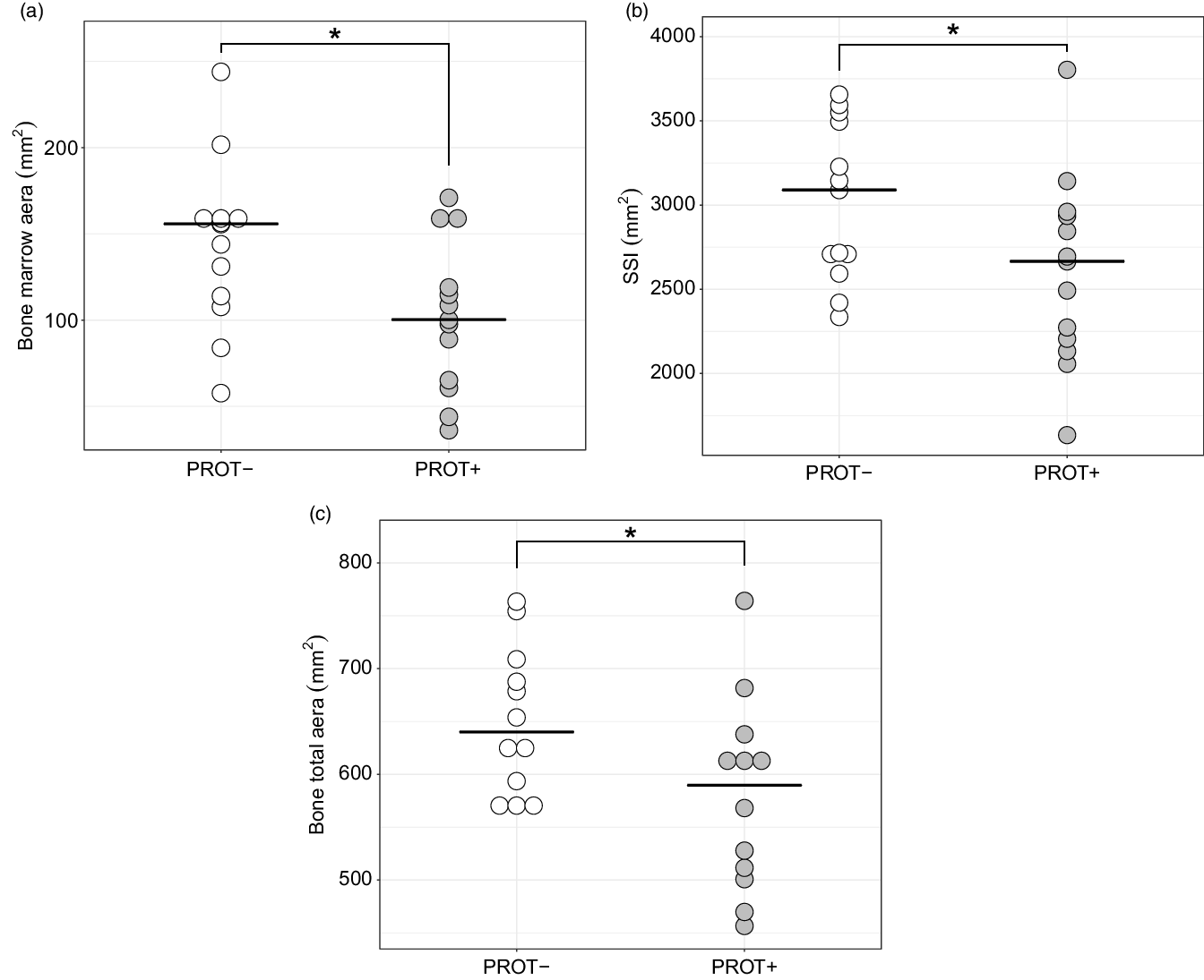
As shown in Fig. 1, marrow area was greater in PROT− than in PROT+ (155 (114–159) v. 100 (65·3–119) mm2; P = 0·049). Bone compressive strength was significantly stronger in the PROT− group than in the PROT+ group (3090 (2709–3496) v. 2666 (2207–2936) mm2; P = 0·048). The PROT− group displayed a higher total bone area compared with the PROT+ group (626 (574–688) v. 568 (501–615) mm2; P = 0·045). No other difference in bone architecture or bone density was found.

Fig. 1 (a) Bone marrow area (mm2), (b) bone compressive strength (SSI, mm2) and (c) bone total area (mm2), according to protein intake (PROT−: < 1 g/kg per d v. PROT+: > 1·2 g/kg per d) among dynapenic-obese older people. *P-value < 0·05
Muscle strength and power
Absolute and relative muscle strength and muscle power were comparable between the two groups (Table 1).
Functional and aerobic capacities
No difference between groups was found for functional and aerobic capacities (Table 1).
Discussion
Despite the low statistical power of the current study, the results suggest that a lower protein intake, but higher than RDA, protects more bone architecture but does not influence bone density among dynapenic-obese older people. The heterogeneity of the population (i.e., very older adults aged 85 years or older(Reference Granic, Mendonca and Sayer18) v. young older adults in the present study), as well as the type of population (malnourished, frail or osteoporotic(Reference Durosier-Izart, Biver and Merminod2) v. healthy adult in the present study) and the difference in study design (i.e., position paper(Reference Bauer, Biolo and Cederholm5), longitudinal study(Reference Granic, Mendonca and Sayer18) v. cross-sectional analysis in the present study) can explain the discrepancies between our conclusion and those from others. Another explanation is that our sample included men and women, whereas Scott et al. showed that only dynapenic-obese men and not women presented more risk of bone deterioration than others(Reference Scott, Blizzard and Fell19). Nevertheless, we cannot investigate these hypotheses since our sample is too small. Finally, the bone health status of our population (without osteoporosis) could have influenced the results of this research. Some limitations are to be emphasised and can explain our conclusion. First, there is a risk of false positive because of the large number of bivariable comparisons performed. Then, the design of the study (i.e., cross-sectional study) does not allow us to establish causal links and the sample size also limits the external validation of the results. Others limitations are the lack of accuracy of the 3-d food diary method, and the fact that confounding variables could not be adjusted. Finally, confounding factors were not taken into account in the analysis, such as age, sex, and in the case of women, time elapsed since menopause and hormonal replacement.
In conclusion, in non-osteoporotic dynapenic-obese young older adults, a lower protein intake seems to be associated with bone sizes, which influence bone strength, but do not influence bone density.
Acknowledgements
Acknowledgements: The authors would like to thank all study participants. Financial support: F.B. is funded by IRSC and FRQS post-doctoral fellowships, A.B. is funded by IRSC and FRQS master fellowships. M.A.-L. is recipient of Chercheur Boursier salary awards from the FRQS. This work was supported by grants from the FRQS, the Réseau Québécois de Recherche sur le Vieillissement, a thematic network funded by FRQS and the Groupe de Recherche en Activité Physique Adaptée – Université du Québec à Montréal. Conflict of interest: The authors declare that they have no conflict of interest. Authorship: M.A.L. and P.N. conceived the original idea. E.P. and A.B. collected the data. F.B. and P.N. performed the statistical analyses. F.B. wrote the manuscript. M.A.L., P.N., E.V. and A.B. revised the manuscript. M.A.L. and P.N. supervised the project. Ethics of human subject participation: The current study was conducted according to the guidelines laid down in the Declaration of Helsinki and all procedures involving research study participants were approved by the Ethics Committee of the Université du Québec à Montréal. Written informed consent was obtained from all subjects/patients.


