Introduction
Posttraumatic stress disorder (PTSD) is highly prevalent in Iraq and Afghanistan-era veterans (Fulton et al., Reference Fulton, Calhoun, Wagner, Schry, Hair, Feeling and Beckham2015) and is associated with adverse outcomes including disability, unemployment, reduced work productivity, and poorer quality of life and physical health (Miloyan, Bulley, Bandeen-Roche, Eaton, & Gonçalves-Bradley, Reference Miloyan, Bulley, Bandeen-Roche, Eaton and Gonçalves-Bradley2016; Schnurr, Lunney, Bovin, & Marx, Reference Schnurr, Lunney, Bovin and Marx2009; Seal et al., Reference Seal, Cohen, Waldrop, Cohen, Maguen and Ren2011; Tanielian & Jaycox, Reference Tanielian and Jaycox2008; Zivin et al., Reference Zivin, Bohnert, Mezuk, Ilgen, Welsh, Ratliff and Kilbourne2011). Trauma-focused cognitive behavioral therapy (CBT), including cognitive processing therapy (CPT), is one of the most effective treatments for PTSD (Bisson, Roberts, Andrew, Cooper, & Lewis, Reference Bisson, Roberts, Andrew, Cooper and Lewis2013; Lee et al., Reference Lee, Schnitzlein, Wolf, Vythilingam, Rasmusson and Hoge2016; Watts et al., Reference Watts, Schnurr, Mayo, Young-Xu, Weeks and Friedman2013). The efficacy and effectiveness of CPT has been demonstrated in a wide range of traumatized populations (Chard, Reference Chard2005; Monson et al., Reference Monson, Schnurr, Resick, Friedman, Young-Xu and Stevens2006; Resick, Nishith, Weaver, Astin, & Feuer, Reference Resick, Nishith, Weaver, Astin and Feuer2002). Yet, a large portion of veterans with PTSD drop out of treatment prematurely or do not adequately respond to treatment (Bradley, Greene, Russ, Dutra, & Westen, Reference Bradley, Greene, Russ, Dutra and Westen2005; Kehle-Forbes, Meis, Spoont, & Polusny, Reference Kehle-Forbes, Meis, Spoont and Polusny2016; Schottenbauer, Glass, Arnkoff, Tendick, & Gray, Reference Schottenbauer, Glass, Arnkoff, Tendick and Gray2008). One way to improve outcomes of PTSD treatment is to identify predictors of response, including biomarkers that may inform treatment mechanisms, with the ultimate goal of matching individuals to treatments where they are most likely to benefit.
A candidate predictor of PTSD treatment outcomes is executive functioning (EF) – the higher-order processes necessary for self-regulation and goal-directed behavior (Banich, Reference Banich2009; Miyake et al., Reference Miyake, Friedman, Emerson, Witzki, Howerter and Wager2000; Snyder, Miyake, & Hankin, Reference Snyder, Miyake and Hankin2015). Intact EF skills are vital for effective CBT given the cognitive tasks involved in treatment (e.g. self-monitoring, inhibiting distorted thoughts, flexibly generating/evaluating alternative thoughts (Aupperle, Melrose, Stein, & Paulus, Reference Aupperle, Melrose, Stein and Paulus2012; Crocker et al., Reference Crocker, Jurick, Thomas, Keller, Sanderson-Cimino, Boyd and Jak2018; Falconer, Allen, Felmingham, Williams, & Bryant, Reference Falconer, Allen, Felmingham, Williams and Bryant2013)). Specific to PTSD, it has been posited that successful treatment involves activation of EF systems to flexibly engage, contextualize, and integrate new information or memories related to traumatic events (Foa, Steketee, & Rothbaum, Reference Foa, Steketee and Rothbaum1989; Liberzon & Sripada, Reference Liberzon and Sripada2008). However, PTSD is associated with deficits in EF (Aupperle et al., Reference Aupperle, Melrose, Stein and Paulus2012; Bomyea, Amir, & Lang, Reference Bomyea, Amir and Lang2012) and corresponding frontoparietal neural circuitry (Collette, Hogge, Salmon, & Van Der Linden, Reference Collette, Hogge, Salmon and Van Der Linden2006; Owen, McMillan, Laird, & Bullmore, Reference Owen, McMillan, Laird and Bullmore2005), including dysfunction in the amygdala, medial prefrontal cortex, hippocampus, dorsal anterior cingulate cortex (ACC), and insula (Fitzgerald, DiGangi, & Phan, Reference Fitzgerald, DiGangi and Phan2018; Hughes & Shin, Reference Hughes and Shin2011; Patel, Spreng, Shin, & Girard, Reference Patel, Spreng, Shin and Girard2012). Cognitive elements of treatment may thus be particularly challenging for a subset of those with PTSD who demonstrated pronounced executive dysfunction. Indeed, worse baseline EF has been associated with poorer response to CBT in several disorders (D'Alcante et al., Reference D'Alcante, Diniz, Fossaluza, Batistuzzo, Lopes, Shavitt and Hoexter2012; Garety et al., Reference Garety, Fowler, Kuipers, Freeman, Dunn, Bebbington and Jones1997; Granholm et al., Reference Granholm, McQuaid, Link, Fish, Patterson and Jeste2008; Mohlman & Gorman, Reference Mohlman and Gorman2005), including CPT for veterans with PTSD (Crocker et al., Reference Crocker, Jurick, Thomas, Keller, Sanderson-Cimino, Boyd and Jak2018, Reference Crocker, Sullan, Jurick, Thomas, Davey, Hoffman and Jak2023). Studies also reveal that functioning of frontoparietal substrates relevant to EF predict treatment response in trauma-focused CBT (Bryant et al., Reference Bryant, Williamson, Erlinger, Felmingham, Malhi, Hinton and Korgaonkar2021; Falconer et al., Reference Falconer, Allen, Felmingham, Williams and Bryant2013; van Rooij, Geuze, Kennis, Rademaker, & Vink, Reference van Rooij, Geuze, Kennis, Rademaker and Vink2015). Taken together, this literature suggests that EF skills may impact clinical outcomes in PTSD treatment, and that EF skills could be an intervention target that is addressed prior to standard PTSD treatment in order to bolster clinical response.
A growing body of literature on computer-based cognitive training, whereby individuals complete repetitive exercises of a specific cognitive skill, demonstrates its promise for improving EF performance (Basak, Boot, Voss, & Kramer, Reference Basak, Boot, Voss and Kramer2008; Nouchi et al., Reference Nouchi, Taki, Takeuchi, Hashizume, Nozawa, Kambara and Kawashima2013; Owen et al., Reference Owen, Hampshire, Grahn, Stenton, Dajani, Burns and Ballard2010). While EF is an umbrella term encompassing a multitude of complex cognitive functions, the most common division of EF is comprised of three core facets, including response inhibition, updating and monitoring of working memory, and mental set shifting (Miyake et al., Reference Miyake, Friedman, Emerson, Witzki, Howerter and Wager2000; Miyake & Friedman, Reference Miyake and Friedman2012). Consequently, EF training programs often incorporate exercises which target these specific EF comsponents. BrainHQ (Posit Science) is a cognitive training program available in an easily accessible online platform that shows efficacy in terms of mental health symptom reduction and/or improved cognition in a range of conditions, including attention-deficit/hyperactivity disorder (Mishra, Sagar, Joseph, Gazzaley, & Merzenich, Reference Mishra, Sagar, Joseph, Gazzaley and Merzenich2016), traumatic brain injury (Mahncke et al., Reference Mahncke, DeGutis, Levin, Newsome, Bell, Grills and Merzenich2021; Voelbel, Lindsey, Mercuri, Bushnik, & Rath, Reference Voelbel, Lindsey, Mercuri, Bushnik and Rath2021), unipolar and bipolar depression (Hagen et al., Reference Hagen, Lau, Joormann, Småstuen, Landrø and Stubberud2020; Lewandowski et al., Reference Lewandowski, Sperry, Cohen, Norris, Fitzmaurice, Ongur and Keshavan2017; Morimoto et al., Reference Morimoto, Altizer, Gunning, Hu, Liu, Cote and Alexopoulos2020), substance use disorders (Bell et al., Reference Bell, Muppala, Weinstein, Ciosek, Pittman, Petrakis and Fiszdon2020; Bell, Vissicchio, & Weinstein, Reference Bell, Vissicchio and Weinstein2016), and mild cognitive impairment (Harvey, Zayas-Bazan, Tibiriçá, Kallestrup, & Czaja, Reference Harvey, Zayas-Bazan, Tibiriçá, Kallestrup and Czaja2022; Levy et al., Reference Levy, Smith, De Wit, DeFeis, Ying, Amofa and Chandler2022). Extant literature demonstrates changes in brain activation and connectivity, including frontoparietal regions, following training with BrainHQ exercises (Chen, Turnbull, Cole, Zhang, & Lin, Reference Chen, Turnbull, Cole, Zhang and Lin2022; Koshiyama et al., Reference Koshiyama, Miyakoshi, Thomas, Joshi, Molina, Tanaka-Koshiyama and Light2020; Lindsey et al., Reference Lindsey, Lazar, Mercuri, Rath, Bushnik, Flanagan and Voelbel2022; McEwen et al., Reference McEwen, Jarrahi, Ventura, Subotnik, Nguyen, Woo and Nuechterlein2023). This research supports a plasticity model of cognitive training, whereby underlying neurocircuitry changes occurring as a result of repeated practice contribute to training-related behavioral performance and clinical gains (Klingberg, Reference Klingberg2010). Taken together, these findings collectively imply that computerized EF training via programs like BrainHQ hold promise for strengthening EF and its associated neural networks. Thus, BrainHQ EF training modules could be viable adjunctive components to PTSD intervention that may enable veterans to fully engage in and benefit from treatment.
However, addressing EF in PTSD treatment may not be a universally helpful adjunctive approach. Cognitive training for EF is mechanistically specific in that it is designed to target a particular set of thinking skills that are both deficient and etiologically involved in symptoms in a subset of individuals. Thus, EF training would likely be of greatest benefit to those with EF deficits. Consistent with this proposal, a recent meta-analysis examining the relationship between baseline cognitive abilities and cognitive training outcomes found that those with weaker baseline EF gained more from cognitive training than those who were more proficient initially (Traut, Guild, & Munakata, Reference Traut, Guild and Munakata2021). However, the extent to which initial neurocognitive deficits relate to interventions that combine EF training with standard psychotherapy has not been well studied to date.
In the current study, we sought to determine the extent to which the neurocognitive substrates of EF predicted clinical response to a novel cognitively enhanced CPT program. We conducted a randomized controlled trial where veterans were randomized to either computerized EF training plus CPT (CEFT + CPT) or placebo training plus CPT (PT + CPT). Prior to engaging in treatment, participants completed a baseline functional magnetic resonance imaging (fMRI) scan in which neural activity during a working memory task was assessed. Given that CEFT + CPT was a targeted intervention to ameliorate specific EF deficits prior to CPT, we expected that those with poorer EF neural activity at baseline would experience greater symptom improvement in this group. Specifically, we predicted that lower baseline neural activity in frontoparietal regions would differentially relate to clinical outcomes in CEFT + CPT v. PT + CPT, such that individuals with reduced baseline EF neural activity would derive greater benefit from CEFT + CPT v. PT + CPT.
Methods
Participants and procedures
The current study consisted of 60 veterans with PTSD who participated in a clinical trial comparing EF training plus CPT (CEFT + CPT; n = 35) to placebo training plus CPT (PT + CPT; n = 25; clinicaltrials.gov identifier NCT03260127) from 2018 to 2022. The sample included those who completed a 2-run working memory task while undergoing functional neuroimaging as part of the parent trial. Veteran participants were recruited primarily through VA San Diego Healthcare System (VASDHS) clinical referrals, in addition to other VASDHS research studies. Potential participants completed a phone screen, then completed a clinical assessment including written informed consent (in accordance with the VASDHS Institutional Review Board, which approved the study) to determine eligibility for the trial. The authors assert that all procedures contributing to this work comply with the ethical standards of the relevant national and institutional committees on human experimentation and with the Helsinki Declaration of 1975, as revised in 2008. Clinical raters and study therapists were blinded to condition.
Participants were included in the clinical trial if they (1) were Iraq/Afghanistan-era veterans enrolled at VASDHS, (2) were aged 18–55, (3) had a current PTSD diagnosis, (4) endorsed subjective cognitive complaints, (5) had no pending psychotropic medication changes (changes in specific medication or dosage in the previous 6 weeks or plans to change in the next month), (6) were English-speaking, and (7) had regular access to a computer to complete EF training. PTSD diagnosis was determined by administration of the Clinician-Administered PTSD Scale for DSM-5 (CAPS-5; Weathers et al., Reference Weathers, Bovin, Lee, Sloan, Schnurr, Kaloupek and Marx2018) and cognitive complaints were assessed via phone screening questions that were based on the Neurobehavioral Symptom Questionnaire cognitive items (Cicerone & Kalmar, Reference Cicerone and Kalmar1995). Exclusion criteria were (1) active substance use disorder in the last month, (2) suicidal intent or attempt within the last month, (3) schizophrenia, psychotic disorder, and/or bipolar disorder, (4) dementia, (5) premorbid IQ below 70, (6) participation in other concurrent PTSD intervention studies, (7) previous completion of more than four CPT sessions, (8) history of a documented neurological disorder (e.g. Parkinson's disease, multiple sclerosis, epilepsy), and (9) moderate-to-severe traumatic brain injury (TBI) according to the Veterans Affairs/Department of Defense (VA/DoD) criteria (2016). While contraindications to MRI were not exclusionary for the parent study, the current study only included those who completed the MRI scan. Of the 82 participants who were eligible, consented, and enrolled in the parent clinical trial, 64 completed the fMRI task. Four participants were excluded due to data quality issues, leaving a total of 60 participants in the current study.
Treatment randomization was stratified by mild TBI status to ensure balanced TBI history across treatment arms. Participants completed six weeks of computerized training (CEFT or PT) and then 12 sessions of standard CPT. Prior to the start of the COVID-19 pandemic in March 2020, clinical assessments and therapy sessions were completed in-person. In order to minimize risk of COVID-19 exposure, all assessments were split into two sessions during the pandemic – one completed in-person and one virtually. From March 2020 onward, all CPT sessions were completed virtually using VA telehealth software. Participants received compensation for their participation.
Interventions
Computerized training
Both the active and control (‘placebo’) computerized training conditions were self-administered via BrainHQ by Posit Science (www.brainhq.com) and consisted of online training on a personal computer for 30 min a day, five times a week for six weeks. Study staff monitored training usage and progress through a secure BrainHQ portal and was in weekly contact with participants to check on their progress, answer questions, and address technical issues. The weekly check-in also served the purpose of keeping participants engaged and motivated. In addition, participants were given a $30 bonus as an incentive for completing at least 80% of the cognitive training. In the current sample, 18 participants in the PT-CPT group received the bonus and 10 in the CEFT-CPT group received the bonus. On average, participants in the PT-CPT group completed 477.35 (s.d. = 281.47) minutes, while those in the CEFT-CPT group completed 366.31(s.d. = 285.16) minutes, t(80) = 1.77, p = 0.08.
Each condition consisted of six training exercises. In the CEFT condition, the six cognitive training exercises targeted executive function processes of inhibition, shifting, and updating of working memory. The exercises were adaptive on a trial-by-trial basis based on an individual's performance. See Supplemental Materials (S1) for descriptions of each of the exercises. Participants in the PT control condition completed six exercises: crossword puzzles, sudoku, connect four, gems swap, maze races, and word search. These exercises were designed to control for nonspecific engagement and motivation of computer games; however, these games were selected to minimize the demands on executive functioning and did not provide the intensive, adaptive training of specific executive functions that the active condition provided.
Cognitive processing therapy
CPT is a gold-standard (trauma-focused), evidence-based treatment for PTSD (Resick et al., Reference Resick, Wachen, Dondanville, Pruiksma, Yarvis and Peterson2017). CPT is a 12-session protocol that involves identifying stuck points (i.e. cognitive distortions) and engaging in cognitive restructuring. Patients learn to challenge and modify stuck points related to traumatic event(s), as well as the themes of safety, trust, power and control, self-esteem, and intimacy. Participants completed CPT sessions 1–2 times a week depending on their schedule. CPT was provided by doctoral-level psychologists who completed formal VA CPT training and certification.
Clinical outcome measure
The primary clinical outcome of interest was the PTSD Checklist for DSM-5 (PCL-5; Blevins et al., Reference Blevins, Weathers, Davis, Witte and Domino2015), past week version, which was measured at 15 timepoints: baseline, after completion of the six weeks of computerized training (CEFT or PT), at each treatment session, and post-CPT.
fMRI task
All participants performed an N-Back working memory task at baseline. In the task, participants monitored a continuous stream of stimuli (pseudowords) and were instructed to respond each time an item was repeated from N before. Each participant completed two N-back runs during fMRI, each consisting of three blocks of trials (i.e. 1-, 2-, or 3-back load conditions). Blocks were counterbalanced over runs. Pseudowords were presented in white font on a black background for 2500 ms with a 500 ms inter-item interval. The timing was constrained so that each block would last exactly 60 s. Blocks were separated by 20 s intervals (with a fixation cross; see Thomas, Duffy, Swerdlow, Light, and Brown (Reference Thomas, Duffy, Swerdlow, Light and Brown2022) for task details) and data were collected over two runs. The primary contrast of interest was 3-back > 1-back.
fMRI data acquisition
Functional imaging was performed with blood-oxygen-level-dependent (BOLD) multiband whole-brain fMRI on a 3.0 Tesla GE 750 (General Electric Healthcare; Waukesha, WI) with a 32-channel head coil. Functional data were acquired using a gradient-echo echo planar imaging (EPI) sequence (TR = 800 ms, TE = 37 ms, flip angle = 52°, acquisition matrix 104 × 104, 2-mm slice thickness, no gaps, 72 ascending axial slices, multiband factor = 6, and 309 volumes per run (with the first 12 removed to achieve steady state, 2 mm isotropic voxels)). A high-resolution T1-weighted structural MPRAGE scan (TR = 8.1 ms, TE = 3.54 ms, flip angle = 8°, thickness = 0.8, no gaps, matrix = 320 mm × 320 mm, voxel size = 0.8 × 0.5 × 0.5 mm) was obtained for co-registration to the functional data and normalization.
fMRI data preprocessing
All imaging analyses were conducted using AFNI version AFNI_22.0.15 (Cox, Reference Cox1996). Preprocessing (afni.proc.py) included despiking, co-registration, spatial smoothing with a 6-mm full-width half-maximum smoothing kernel and warping to Montreal Neurologic Institute (MNI) space. Four task response regressors were generated at the individual subject level for the task reflecting trial type (1-back, 2-back, 3-back) and a high-low contrast (3-back > 1-back). The regression included censoring of large motion (≥0.3 mm) between successive time points. Prior to analysis, visual inspection for alignment and percent of censored data (<20%) was conducted, resulting in removal of four subjects who failed quality control checks. Analysis of whole-brain task effects in the sample can be found in the Supplemental Materials (S2).
Data analysis plan
A region-of-interest (ROI) approach was used to examine the relationship between baseline neural functioning during a working memory task and change in PTSD symptoms over the course of treatment. ROIs were created using Neurosynth (Neurosynth.org; see Supplemental Materials Table S2 and Figure S2 for regions). Parameter estimates of the contrast of interest (3-back > 1-back) were extracted from each ROI and entered into separate linear mixed effects models using restricted maximum likelihood estimation procedures. Each model contained predictors of mean-centered extracted neural activity to the contrast, treatment group, time (corresponding to weeks from baseline, centered at baseline), and their respective interaction terms with subject included as a random effect. We specifically examined the extent to which neural activity at baseline was a prescriptive predictor (Fournier et al., Reference Fournier, DeRubeis, Shelton, Hollon, Amsterdam and Gallop2009; Kraemer, Wilson, Fairburn, & Agras, Reference Kraemer, Wilson, Fairburn and Agras2002), that is, whether there was a significant neural activity by time by treatment condition interaction for each ROI. False discovery rate (FDR) was used to correct for multiple comparisons. In cases where a prescriptive predictor model was significant, we conducted a follow-up analysis split by treatment group to clarify the source of the effects (i.e. a neural activity × time effect separately within each group).
Results
Demographic, clinical, and behavioral characteristics
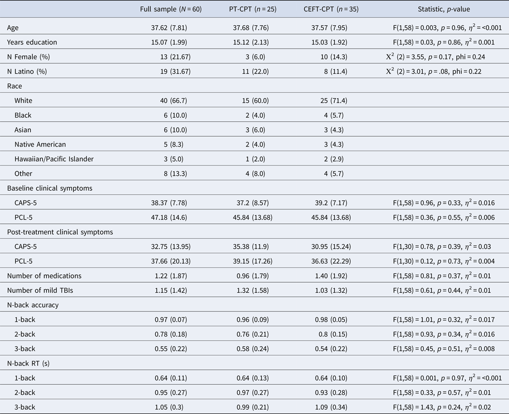
Table 1 presents demographic and clinical characteristics of the sample. The average age of the sample was 37.62 years (range of 23–53). Mean pre-treatment PCL-5 was 47.18 (s.d. = 14.60), indicating elevated baseline severity of PTSD. Mean post-treatment PCL-5 was 37.66 (s.d. = 20.13). Results revealed a significant decrease in PTSD symptoms over time across groups, B = −0.54, s.e. = .07, p < 0.001 (full results comparing treatment effects can be found in Crocker, Reference Crocker2025). Table 1 also presents average reaction time and accuracy for each trial type (1-, 2-, and 3-back). In the full sample, comparing behavioral performance across N-back trial types revealed a significant decrease in accuracy, F(2118) = 113.0, p < 0.001, and a significant increase in reaction time, F(2118) = 56.10, p < 0.001, as task difficulty increased. With regard to prior treatment for PTSD in the full sample, two participants completed minimal sessions of prior CPT (four or fewer sessions), five had completed sessions of prolonged exposure, four underwent other cognitive behavioral therapy, 11 experienced supportive therapy, and nine reported other or unknown therapies. In the PT-CPT arm, the mean length of time participants reported experiencing PTSD symptoms was 106.1 months (s.d. = 62.93), while the mean time was 137.4 months (s.d. = 91.00) for participants in the CEFT-CPT arm. There was no difference between groups in terms of time experiencing PTSD symptoms, F(1,58) = 2.20, p = 0.14.
Table 1. Clinical and demographic characteristics of full sample, in addition to N-back behavioral task performance variables; all values are means unless otherwise indicated and standard deviations are in parentheses

Note: All clinical measures represent total scores.
PT-CPT, placebo training plus cognitive processing therapy treatment arm; CEFT-CPT, computerized executive functioning training plus cognitive processing therapy treatment arm; CAPS-5, Clinician Administered PTSD Scale for DSM-5 – Past Month; PCL-5, PTSD Checklist for DSM-5 – Past Week; TBI, traumatic brain injury.
Relationship between neural activity at baseline and PTSD symptom change
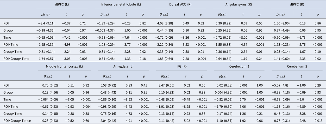
Results of longitudinal models predicting PTSD symptom change from neural activity at baseline are presented in Table 2. Results of the model revealed neural activity in the left dorsolateral PFC (dlPFC), right dorsal ACC, left amygdala, and right inferior frontal gyrus (IFG). For each of these ROIs, individuals with relatively greater neural response to 3-back v. 1-back showed greater PTSD symptom improvement trajectories in CEFT + CPT v. PT + CPT, while individuals with relatively less neural response to the contrast showed similar PTSD symptom change irrespective of treatment condition (see Fig. 1). No other ROIs reached statistical significance after adjusting for multiple comparisons (ps > 0.02).
Table 2. Relationships between baseline neural activity during a working memory task and change in PTSD symptoms over treatment

Note: Group represents treatment group, CEFT-CPT v. PT-CPT; Cerebellum 1 and 2 were two distinct regions identified within the cerebellum.
ROI, region of interest; dlPFC, dorsolateral prefrontal cortex; ACC, anterior cingulate cortex; IFG, inferior frontal gyrus.

Figure 1. Neural activation in (a) dlPFC (L), (b) dACC, (c) amygdala (L), and (d) IFG (R) to 3 back >1 back was a prognostic predictor in treatment condition model (CEFT-CPT v. PT-CPT): Estimated PCL-5 scores from baseline to post-treatment sessions at 1 s.d. above and below the mean of extracted BOLD signal. ROIs reflect anatomical regions based on the Neurosynth meta-analytic mask.
Follow-up analyses revealed that there was a significant association between neural effects and clinical outcome in the CEFT-CPT group, but not the PT-CPT group, in the left dlPFC (CEFT-CPT: β = −1.99 (0.42), p < 0.001; PT-CPT: β = −0.14 (0.40), p = 0.72), right dorsal ACC (CEFT-CPT: β = −2.24 (0.36), p < 0.001; PT-CPT: β = −0.29 (0.51), p = .58), and right IFG (CEFT-CPT: β = −1.93 (0.24), p < 0.001; PT-CPT: β = −.27 (0.34), p = 0.44). The left amygdala ROI was associated with greater symptom reduction in both groups (CEFT-CPT: β = −1.03 (0.31), p < 0.001; PT-CPT: β = −0.09 (0.11), p < 0.001). To assist in making inferences about the directionality of neural effects relative to performance, we conducted Spearman's correlations between N-back task accuracy with the significant ROIs from our previous models. Results revealed that greater activation in the right dorsal ACC, r = 0.24, p < 0.005, and right IFG, r = 0.21, p < 0.017, was associated with better accuracy, suggesting that greater activation reflected greater working memory performance.
Discussion
The current study sought to examine relationships between baseline neural correlates of working memory and PTSD symptom change in veterans enrolled in a randomized controlled trial testing a novel treatment combining EF training with CPT (CEFT + CPT v. PT + CPT). We evaluated the extent to which neural activation was a prescriptive predictor. That is, we determined if baseline neural activity differentially related to clinical PTSD outcomes measured using weekly PCL-5 scores administered throughout treatment. We found that frontoparietal regions were differentially predictive of outcomes depending on treatment group. Contrary to study hypotheses, greater (rather than less) activation of regions implicated in working memory (i.e. dlPFC, dorsal ACC, amygdala, and IFG) was associated with better PTSD symptom improvement trajectories in CEFT + CPT. In contrast, neural activity did not show statistically significant associations with PTSD symptom change in PT-CPT.
Results suggest that activity of frontal regions implicated in working memory may serve as an important biomarker of response to a cognitively enhanced treatment in veterans with PTSD. Findings from the current study are consistent with a large body of work that highlights the role of a frontoparietal network in working memory and EF (Collette et al., Reference Collette, Hogge, Salmon and Van Der Linden2006; Owen et al., Reference Owen, McMillan, Laird and Bullmore2005). This network, in which the dlPFC and IFG are core regulatory components, facilitate ‘cold’ executive skills and processes inclusive of working memory, which regulate the series of decisions involved in the planning and execution of goal-directed behavior (Ward, Reference Ward2019). The dlPFC is responsible for various working memory operations, including attention allocation and task-relevant functions such as the monitoring and manipulation of cognitive representations (Barbey, Koenigs, & Grafman, Reference Barbey, Koenigs and Grafman2013; D'Esposito, Postle, & Rypma, Reference D'Esposito, Postle and Rypma2000), while the right IFG is implicated in cognitive inhibition, a component of executive control defined as the suppression of inappropriate responses (Aron, Robbins, & Poldrack, Reference Aron, Robbins and Poldrack2004; Molnar-Szakacs, Iacoboni, Koski, & Mazziotta, Reference Molnar-Szakacs, Iacoboni, Koski and Mazziotta2005), in addition to attentional switching (Cools, Clark, Owen, & Robbins, Reference Cools, Clark, Owen and Robbins2002; Dove, Pollmann, Schubert, Wiggins, & von Cramon, Reference Dove, Pollmann, Schubert, Wiggins and von Cramon2000; Hampshire & Owen, Reference Hampshire and Owen2006). Additionally, the cingulate is a key node within salience and cognitive control networks that signals the importance of external stimuli (Niendam et al., Reference Niendam, Laird, Ray, Dean, Glahn and Carter2012). The cingulate cortex (i.e. dorsal ACC) is theorized to play an integrative role across multiple brain systems in the service of cost/benefit computations necessary for tenacity during demanding tasks (Touroutoglou, Andreano, Dickerson, & Barrett, Reference Touroutoglou, Andreano, Dickerson and Barrett2020), in addition to aiding in conflict monitoring and action selection and execution (Stevens, Hurley, & Taber, Reference Stevens, Hurley and Taber2011; Vassena, Holroyd, & Alexander, Reference Vassena, Holroyd and Alexander2017; Vogt, Reference Vogt and Vogt2019). Our correlational findings, alongside prior data examining low spans on the N-back task (Lamichhane, Westbrook, Cole, & Braver, Reference Lamichhane, Westbrook, Cole and Braver2020), suggest that greater activity across these regions supports working memory ability.
The present findings expand prior literature that demonstrates frontoparietal functioning relates to PTSD treatment outcomes (Bryant et al., Reference Bryant, Williamson, Erlinger, Felmingham, Malhi, Hinton and Korgaonkar2021; Falconer et al., Reference Falconer, Allen, Felmingham, Williams and Bryant2013; van Rooij et al., Reference van Rooij, Geuze, Kennis, Rademaker and Vink2015). Several methodological features differ between our study and this earlier work, including treatment modality (CPT v. other trauma focused therapy), use of a comparator group, and task type (working memory v. response inhibition); these features may account for differences in findings. Results from the present study indicated that veterans who had relatively higher baseline neural response to working memory processing fared better in treatment – but this effect was specific to those receiving cognitive training in conjunction with CPT rather than placebo training paired with standard CPT. These findings align with a capitalization model of treatment response, whereby a given treatment is best matched to an individual when it builds upon their strengths (i.e. improving cognitive skill in those with greater baseline ability [Cheavens, Strunk, Lazarus, & Goldstein, Reference Cheavens, Strunk, Lazarus and Goldstein2012]) rather than attempting to target a relative weakness.
It has been posited that successful PTSD treatment involves activation of EF systems to flexibly engage, contextualize, and integrate new information or memories related to traumatic events (Foa et al., Reference Foa, Steketee and Rothbaum1989; Liberzon & Sripada, Reference Liberzon and Sripada2008). For instance, CPT includes skills such as self-monitoring to identify trauma-related beliefs, inhibiting or disengaging from distorted thoughts and evaluating them, and flexibly generating alternative thoughts. These skills likely draw upon EF and the coordinated activity of regions that support these functions, including frontoparietal areas. At its core, CPT teaches clients to increase and refine the use of EF to manage emotional distress. Consequently, those with better baseline neurocognitive ability may have benefited the most from training in terms of obtaining the greatest EF gains, resulting in better downstream effects during treatment on processes linked to cognitive skills (e.g. better cognitive flexibility to modify maladaptive thoughts or inhibit negative thinking [Bomyea & Lang, Reference Bomyea and Lang2015] and improved emotion regulation [Schmeichel, Volokhov, & Demaree, Reference Schmeichel, Volokhov and Demaree2008]). Put differently, those with better EF brain function may capitalize on that activation during computerized EF training, priming those veterans to effectively employ EF skills in treatment and effectively reduce PTSD symptoms. Further research will be needed to clarify mechanisms by which baseline EF activation confers clinical benefit in this type of intervention.
This study has a number of limitations. First, the sample size was relatively small and consisted primarily of male Iraq and Afghanistan-era veterans. Additional research is needed in a larger and more generalizable sample to extend findings to female veterans, veterans of other eras, and to non-Veteran populations. Veterans did not complete posttreatment imaging sessions and, therefore, we can only speak to the effect of initial neural activity and cannot make inferences about the impact of treatment on working memory brain functions. Neuroimaging prediction analyses have inherent limitations, including in some cases low feasibility for utilization clinically given the high cost and participant burden. However, proxies of neural functioning (based on indirect physiological, behavioral, neuropsychological, and ecological momentary assessment measures, among others) could ultimately be helpful to identify those who would benefit most from particular treatments once neural predictors are well validated. Regarding mechanisms, we are unable to determine which aspects of the computerized executive function training translate to better treatment outcomes and, thus, future research is needed to identify the specific mechanisms by which those with greater activation is working memory-related brain regions derive benefit from cognitive training. Moreover, we utilized a working memory task, which evaluates one of three core facets of EF (Miyake et al., Reference Miyake, Friedman, Emerson, Witzki, Howerter and Wager2000; Miyake & Friedman, Reference Miyake and Friedman2012); as a result, it is unclear whether similar findings would emerge for other EF tasks (e.g. inhibition).
Despite limitations, the present study is among the first to explore neural predictors of treatment-related PTSD symptom change – specifically, neural activity of a working memory task at baseline predicting clinical outcomes following a novel cognitive training intervention. Clinically, findings have the potential to inform precision medicine and increase our understanding of treatment mechanisms. Veterans with greater frontal brain activity related to working memory at baseline may be better able to utilize EF training, which could have had downstream positive impacts on psychotherapy effectiveness. Future research is needed to elucidate the mechanisms by which activity in brain regions implicated in working memory contribute to improved treatment response for those who complete computerized executive function training prior to CPT. Moreover, future studies should identify whether neural activity associated with other EF components (i.e. inhibition) also predict treatment response.
Supplementary material
The supplementary material for this article can be found at https://doi.org/10.1017/S0033291724003106.
Acknowledgments
The authors sincerely thank Michael P. Thomas for his contributions to the N-back task.
Funding statement
Funding for the clinical trial was supported by a Career Development Award from the VA Rehabilitation Research & Development Service (LC, grant number IK2 RX002459). The funding agencies had no role in data collection, analysis, or manuscript development. No potential conflicts of interest were reported by coauthors. The views, opinions, and/or findings contained in this article are those of the authors and should not be construed as an official Veterans Affairs or Department of Defense position, policy, or decision unless so designated by other official documentation.
Competing interests
The author(s) declare none.


