Hostname: page-component-745bb68f8f-s22k5 Total loading time: 0 Render date: 2025-01-14T22:35:22.676Z Has data issue: false hasContentIssue false

Biocompatible methionine-capped CdS/ZnS quantum dots for live cell nucleus imaging

Published online by Cambridge University Press:  29 January 2019

S. Kanagasubbulakshmi
Affiliation:
DRDO—BU Center for Life Sciences, Bharathiar University Campus, Coimbatore, 641046, India
I. Gowtham
Affiliation:
DRDO—BU Center for Life Sciences, Bharathiar University Campus, Coimbatore, 641046, India
K. Kadirvelu*
Affiliation:
DRDO—BU Center for Life Sciences, Bharathiar University Campus, Coimbatore, 641046, India
K. Archana
Affiliation:
Department of Biotechnology, PSG College of Arts & Science, Coimbatore, 641014, India
*
Address all correspondence to K. Kadirvelu at kadirvelukrishna@yahoo.com
Get access

Abstract

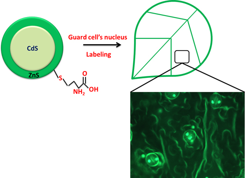
CdS/ZnS core shell quantum dots (QDs) were synthesized and functionalized by methionine and characterized by standard techniques. The prior QD-based phytotoxicity assay was helpful to find out the maximum tolerant level of the plant cells. The successful transport and phytotoxic mechanism of QDs were elaborated in detail. Methionine functionalities on the QDs were helpful in specific binding of QDs with the nucleus of stomata in plant cells. Target specific interaction with the nucleus of stomata cells was a novel breakthrough that can be used in many biologic applications.

Type
Research Letters
Copyright
Copyright © Materials Research Society 2019 

Access options

Get access to the full version of this content by using one of the access options below. (Log in options will check for institutional or personal access. Content may require purchase if you do not have access.)

References

1.Mattoussi, H., Matthew Mauro, J., Goldman, E.R., Anderson, G.P., Sundar, V.C., Mikulec, F.V., and Bawendi, M.G.: Self-assembly of CdS-ZnS quantum dot bioconjugates using an engineered recombinant protein. J. Am. Chem. Soc. 122, 1214212150 (2000).Google Scholar
2.Peng, Z.A. and Peng, X.: Formation of high-quality CdTe, CdSe, and CdS nanocrystals using CdO as precursor. J. Am. Chem. Soc. 123, 183184 (2001).Google Scholar
3.Dabbousi, B.O., Rodriguez-Viejo, J., Mikulec, F.V., Heine, J.R., Mattoussi, H., Ober, R., Jensen, K.F., and Bawendi, M.G.: (CdSe) ZnS core–shell quantum dots: synthesis and characterization of a size series of highly luminescent nanocrystallites. J. Phys. Chem. B. 101, 94639475 (1997).Google Scholar
4.Chan, W.C.W. and Nie, S.: Quantum dot bioconjugates for ultrasensitive nonisotopic detection quantum dot bioconjugates for ultrasensitive nonisotopic detection. Science 281, 20162018 (1998).Google Scholar
5.Bruchez, M., Moronne, M., Gin, P., Weiss, S., and Paul Alivisatos, A.: Semiconductor nanocrystals as fluorescent biological labels. Science 5385, 20132016 (1998).Google Scholar
6.Michalet, X., Pinaud, F.F., Bentolila, L.A., Tsay, J.M., Doose, S.J.J.L., Li, J.J., Sundaresan, G., Wu, A.M., Gambhir, S.S., and Weiss, S.: Quantum dots for live cells, in vivo imaging, and diagnostics. Science 5709, 538544 (2005).Google Scholar
7.Alimohammadi, M., Xu, Y., Wang, D., Biris, A.S., and Khodakovskaya, M.V.: Physiological responses induced in tomato plants by a two-component nanostructural system composed of carbon nanotubes conjugated with quantum dots and its in vivo multimodal detection. Nanotechnology. 29, 295101 (2011).Google Scholar
8.Lidke, D.S., Lidke, K.A., Rieger, B., Jovin, T.M., and Arndt-Jovin, D.J.: Reaching out for signals: filopodia sense EGF and respond by directed retrograde transport of activated receptors. J. Cell Biol. 4, 619626 (2005).Google Scholar
9.Li, W., Zheng, Y., Zhang, H., Liu, Z., Su, W., Chen, S., Liu, Y., Zhuang, J., and Lei, B.: Phytotoxicity, uptake, and translocation of fluorescent carbon dots in mung bean plants. ACS Appl. Mater. Interfaces 8, 1993919945 (2016).Google Scholar
10.Monica, R.C. and Cremonini, R.: Nanoparticles and higher plants. Caryologia 62, 161165 (2009).Google Scholar
11.Nair, R., Poulose, A.C., Nagaoka, Y., Yoshida, Y., Maekawa, T., and Sakthi Kumar, D.: Uptake of FITC labeled silica nanoparticles and quantum dots by rice seedlings: effects on seed germination and their potential as biolabels for plants. J. Fluoresc. 21, 20572068 (2011).Google Scholar
12.Peng, J., Sun, Y., Liu, Q., Yang, Y., Zhou, J., Feng, W., Zhang, X., and Li, F.: Upconversion nanoparticles dramatically promote plant growth without toxicity. Nano Res. 5, 770782 (2012).Google Scholar
13.Koo, Y., Wang, J., Zhang, Q., Zhu, H., Wassim Chehab, E., Colvin, V.L., Alvarez, P.J.J., and Braam, J.: Fluorescence reports intact quantum dot uptake into roots and translocation to leaves of Arabidopsis thaliana and subsequent ingestion by insect herbivores. Environ. Sci. Technol. 49, 626632 (2015).Google Scholar
14.Chakravarty, D., Erande, M.B., and Late, D.J.: Graphene quantum dots as enhanced plant growth regulators: effects on coriander and garlic plants. J. Sci. Food Agric. 95, 27722778 (2015).Google Scholar
15.Thambidurai, M., Muthukumarasamy, N., Agilan, S., Sabari Arul, N., Murugan, N., and Balasundaraprabhu, R.: Structural and optical characterization of Ni-doped CdS quantum dots. J. Mater. Sci. 46, 32003206 (2011).Google Scholar
16.Mirnajafizadeh, F., Wang, F., Reece, P., and Arron Stride, J.: Synthesis of type-II CdSe (S)/Fe2O3 core/shell quantum dots: the effect of shell on the properties of core/shell quantum dots. J. Mater. Sci. 51, 52525258 (2016).Google Scholar
17.Levine, R.L., Mosoni, L., Berlett, B.S., and Stadtman, E.R.: Methionine residues as endogenous antioxidants in proteins. Proc. Natl. Acad. Sci. 93, 1503615040 (1996).Google Scholar
18.Javed Raza, A., Qureshi, S.M.Z., and Khan, M.S.: Synthesis and study of catalytic application of l-methionine protected gold nanoparticles. Appl. Nanosci. 7, 429437 (2017).Google Scholar
19.Jagminas, A., Mikalauskaitė, A., Karabanovas, V, and Vaičiūnienė, J.: Methionine-mediated synthesis of magnetic nanoparticles and functionalization with gold quantum dots for theranostic applications. Beilstein J. Nanotechnol. 8, 1734 (2017).Google Scholar
20.Pazhanivel, T., Nataraj, D., Devarajan, V.P., Mageshwari, V., Senthil, K., and Soundararajan, D.: Improved sensing performance from methionine capped CdTe and CdTe/ZnS quantum dots for the detection of trace amounts of explosive chemicals in liquid media. Anal. Methods 5, 910916 (2013).Google Scholar
21.Lin, D. and Xing, B.: Phytotoxicity of nanoparticles: inhibition of seed germination and root growth. Environ. Pollut. 150, 243250 (2007).Google Scholar
22.Blois, M.S.: Antioxidant determinations by the use of a stable free radical. Nature 181, 11991200 (1958).Google Scholar
23.Zhai, C., Zhang, H., Du, N., Chen, B., Huang, H., Wu, Y., and Yang, D.: One-pot synthesis of biocompatible CdSe/CdS quantum dots and their applications as fluorescent biological labels. Nanoscale Res. Lett. 6, 15 (2011).Google Scholar
24.Li, M., Zhou, H., Zhang, H., Sun, P., Yi, K., Wang, M., Dong, Z., and Xu, S.: Preparation and purification of L-cysteine capped CdTe quantum dots and its self-recovery of degenerate fluorescence. J. Lumin. 130, 19351940 (2010).Google Scholar
25.Gaur, G., Koktysh, D.S., Fleetwood, D.M., Weller, R.A., Reed, R.A., Rogers, B.R., and Weiss, S.M.: Influence of ionizing radiation and the role of thiol ligands on the reversible photodarkening of CdTe/CdS quantum dots. ACS Appl. Mater. Interfaces 8, 78697876 (2016).Google Scholar
26.Thangadurai, P., Balaji, S., and Manoharan, P.T.: Surface modification of CdS quantum dots using thiols—structural and photophysical studies. Nanotechnol. 19, 435708 (2008).Google Scholar
27.Mohammad, F. and Al-Lohedan, H.A.: Toxicity assessment of engineered Mn–ZnS quantum dots in vitro. J. Mater. Sci. 51, 92079216 (2016).Google Scholar
28.Liu, Q., Zhao, Y., Wan, Y., Zheng, J., Zhang, X., Wang, C., Fang, X., and Lin, J.: Study of the inhibitory effect of water-soluble fullerenes on plant growth at the cellular level. ACS Nano 4, 57435748 (2010).Google Scholar
29.Hoshino, A., Fujioka, K., Oku, T., and Suga, M.: Physicochemical properties and cellular toxicity of nanocrystal quantum dots depend on their surface modification. Nano Lett. 4, 21632169 (2004).Google Scholar
30.Xing, R., Chen, X.D., Zhou, Y.F., Zhang, J., Su, Y.Y., Qiu, J.F., Sima, Y.H., Zhang, K.Q., He, Y., and Xu, S.Q.: Targeting and Retention Enhancement of Quantum Dots Decorated with Amino Acids in an Invertebrate Model Organism. Sci. Rep. 6, 110 (2016).Google Scholar
31.Zhao, X., Joo, J., Kim, D., Lee, J., and Kim, J.Y.: Estimation of the Seedling Vigor Index of Sunflowers Treated with Various Heavy Metals. J. Biorem. Biodegrad. 7, 16 (2016).Google Scholar
32.Erofeeva, E.A.: Hormesis and paradoxical effects of wheat seedling (Triticum aestivum L.) parameters upon exposure to different pollutants in a wide range of doses. Dose. Response. 12, 121135 (2014).Google Scholar
33.Yruela, I.: Copper in plants. Brazilian J. Plant Physiol. 1, 145156 (2005).Google Scholar
34.Chibuike, G.U. and Obiora, S.C.: Heavy metal polluted soils: effect on plants and bioremediation methods. Biorem. Meth. 2014, 112 (2014).Google Scholar
35.Müller, F., Houben, A., Barker, P.E., Xiao, Y., Käs, J.A., and Melzer, M.: Quantum dots—a versatile tool in plant science? J. Nanobiotechnol. 4, 5 (2006).Google Scholar
36.Ravindran, S., Kim, S., Martin, R., Lord, E.M., and Ozkan, C.S.: Quantum dots as bio-labels for the localization of a small plant adhesion protein. Nanotechnology 16, 14 (2005).Google Scholar
37.Al-Salim, N., Barraclough, E., and Burgess, E.: Brent Clothier, Markus Deurer, Steve Green, Louise Malone, and Graham Weir: Quantum dot transport in soil, plants, and insects. Sci. Total Environ. 409, 32373248 (2011).Google Scholar
38.McCulloh, K.A., Sperry, J.S., and Adler, F.R.: Water transport in plants obeys Murray's law. Nature 6926, 939 (2003).Google Scholar
39.Libault, M., Tessadori, F., Germann, S., Snijder, B., Fransz, P., and Gaudin, V.: The Arabidopsis LHP1 protein is a component of euchromatin. Planta 222, 910925 (2005).Google Scholar
40.Meierhans, D. and Allemann, R.K.: The N-terminal methionine is a major determinant of the DNA binding specificity of MEF-2C. J. Biol. Chem. 273, 2605226060 (1998).Google Scholar
41.Mgcina, L.S., Dubery, I.A., and Piater, L.A.: Comparative conventional-and quantum dot-labeling strategies for LPS binding site detection in Arabidopsis thaliana mesophyll protoplasts. Front. Plant Sci. 6, 111 (2015).Google Scholar
42.Djikanović, D., Kalauzi, A., Jeremić, M., Xu, J., Mićić, M., Whyte, J.D., Leblanc, R.M., and Radotić, K.: Interaction of the CdSe quantum dots with plant cell walls. Colloids Surf., B 91, 4147 (2012).Google Scholar
43.Ahmed, K.B.A., Ahalya, P., Sengan, M., Kamlekar, R., and Veerappan, A.: Synthesis and characterization of zinc sulfide quantum dots and their interaction with snake gourd (Trichosanthes anguina) seed lectin. Spectrochim. Acta—Part A: Mol. Biomol. Spectrosc. 151, 739745 (2015).Google Scholar
44.Navarro, D.A., Bisson, M.A., and Aga, D.S.: Investigating uptake of water-dispersible CdSe/ZnS quantum dot nanoparticles by Arabidopsis thaliana plants. J. Hazard. Mater., 211–212, 427435 (2012).Google Scholar
Supplementary material: File

Kanagasubbulakshmi et al. supplementary material

Figures S1-S10

Download Kanagasubbulakshmi et al. supplementary material(File)
File 1.2 MB