Hostname: page-component-745bb68f8f-b6zl4 Total loading time: 0 Render date: 2025-01-15T00:04:18.922Z Has data issue: false hasContentIssue false

Stable near-infrared photoluminescence from silicon quantum dot–bovine serum albumin composites

Published online by Cambridge University Press:  11 November 2020
Asuka Inoue
Affiliation:
Department of Electrical and Electronic Engineering, Graduate School of Engineering, Kobe University, Rokkodai, Nada, Kobe, Japan
Hiroshi Sugimoto
Affiliation:
Department of Electrical and Electronic Engineering, Graduate School of Engineering, Kobe University, Rokkodai, Nada, Kobe, Japan
Yozo Sugimoto
Affiliation:
Department of Electrical and Electronic Engineering, Graduate School of Engineering, Kobe University, Rokkodai, Nada, Kobe, Japan
Kensuke Akamatsu
Affiliation:
Department of Nanobiochemistry, Frontiers of Innovative Research in Science and Technology (FIRST), Konan University, Minatojimaminami, Chuo-ku, Kobe, Japan
Marie Hubalek Kalbacova
Affiliation:
Institute of Pathological Physiology, 1st Faculty of Medicine, Charles University, Prague, Czech Republic
Chiaki Ogino
Affiliation:
Department of Chemical Science and Engineering, Graduate School of Engineering, Kobe University, Rokkodai, Nada, Kobe, Japan
Minoru Fujii*
Affiliation:
Department of Electrical and Electronic Engineering, Graduate School of Engineering, Kobe University, Rokkodai, Nada, Kobe, Japan
*
Address all correspondence to Minoru Fujii at fujii@eedept.kobe-u.ac.jp
Get access

Abstract

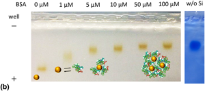
The interaction between negatively charged all-inorganic silicon quantum dots (Si QDs) and bovine serum albumin (BSA) is studied. It is shown that a small difference in the size of Si QDs affects the structure of Si QD–BSA composites significantly. When the diameter of Si QDs is 4 nm, a heterodimer (~20 nm) composed of one Si QD and one BSA molecule is a preferable and stable structure. On the other hand, when the diameter is 7 nm, the size of the composites increases to ~50 nm. The Si QD–BSA composites exhibit stable photoluminescence in the near-infrared range in phosphate-buffered saline.

Type
Research Letters
Copyright
Copyright © The Author(s), 2020, published on behalf of Materials Research Society by Cambridge University Press

Access options

Get access to the full version of this content by using one of the access options below. (Log in options will check for institutional or personal access. Content may require purchase if you do not have access.)

References

Resch-Genger, U., Grabolle, M., Cavaliere-Jaricot, S., Nitschke, R., and Nann, T.: Quantum dots versus organic dyes as fluorescent labels. Nat. Methods 5, 763775 (2008).CrossRefGoogle ScholarPubMed
Wang, Y., Hu, R., Lin, G., Roy, I., and Yong, K.T.: Functionalized quantum dots for biosensing and bioimaging and concerns on toxicity. ACS Appl. Mater. Interfaces 5, 27862799 (2013).10.1021/am302030aCrossRefGoogle ScholarPubMed
Wegner, K.D. and Hildebrandt, N.: Quantum dots: bright and versatile in vitro and in vivo fluorescence imaging biosensors. Chem. Soc. Rev. 44, 47924834 (2015).CrossRefGoogle Scholar
Bruchez, M. Jr., Mario, M., Peter, G., Shimon, W., and Alivisatos, A.P.: Semiconductor nanocrystals as fluorescent biological labels. Science 281, 20132016 (1998).10.1126/science.281.5385.2013CrossRefGoogle ScholarPubMed
Michalet, X.: Quantum dots for live cells, in vivo imaging, and diagnostics. Science 307, 538544 (2005).CrossRefGoogle ScholarPubMed
Derfus, A.M., Chan, W.C.W., and Bhatia, S.N.: Probing the cytotoxicity of semiconductor quantum dots. Nano Lett. 4, 1118 (2004).10.1021/nl0347334CrossRefGoogle ScholarPubMed
Montalti, M., Cantelli, A., and Battistelli, G.: Nanodiamonds and silicon quantum dots: ultrastable and biocompatible luminescent nanoprobes for long-term bioimaging. Chem. Soc. Rev. 44, 48534921 (2015).CrossRefGoogle ScholarPubMed
Farrell, N.O.: Silicon nanoparticles: applications in cell biology and medicine. Int. J. Nanomed. 1, 451472 (2006).10.2147/nano.2006.1.4.451CrossRefGoogle Scholar
Peng, F., Su, Y., Zhong, Y., Fan, C., Lee, S., and He, Y. : Silicon nanomaterials platform for bioimaging, biosensing, and cancer therapy. Acc. Chem. Res. 47, 612623 (2014).CrossRefGoogle ScholarPubMed
Xu, G., Zeng, S., Zhang, B., Swihart, M., Yong, K., and Prasad, P.: New generation cadmium-free quantum dots for biophotonics and nanomedicine. Chem. Rev. 116, 1223412327 (2016).CrossRefGoogle ScholarPubMed
Joo, J., Liu, X., Kotamraju, V., Ruoslahti, E., Nam, Y., and Sailor, M.: Gated luminescence imaging of silicon nanoparticles. ACS Nano 9, 62336241 (2015).10.1021/acsnano.5b01594CrossRefGoogle ScholarPubMed
Kim, D., Kang, J., Wang, T., Ryu, H., Zuidema, J., Joo, J., Kim, M., Huh, Y., Jung, J., Ahn, K., Kim, K., and Sailor, M.: Two-photon in vivo imaging with porous silicon nanoparticles. Adv. Mater. 29, 18 (2017).10.1002/adma.201703309CrossRefGoogle ScholarPubMed
Erogbogbo, F., Yong, K., Roy, I., Xu, G., Prasad, P., and Swihart, M.: Biocompatible luminescent silicon quantum dots for imaging of cancer cells. ACS Nano 2, 873878 (2008).CrossRefGoogle ScholarPubMed
Erogbogbo, F., Yong, K., Roy, I., Hu, R., Law, W., Zhao, W., Ding, H., Wu, F., Kumar, R., Swihart, M., and Prasad, P.: In vivo targeted cancer imaging, sentinel lymph node mapping and biocompatible silicon nanocrystals. ACS Nano 5, 413423 (2011).10.1021/nn1018945CrossRefGoogle ScholarPubMed
Clark, R.J., Dang, M.K.M., and Veinot, J.G.C.: Exploration of organic acid chain length on water-soluble silicon quantum dot surfaces. Langmuir 26, 1565715664 (2010).10.1021/la102983cCrossRefGoogle ScholarPubMed
Wheeler, L.M., Kramer, N.J., and Kortshagen, U.R.: Thermodynamic driving force in the spontaneous formation of inorganic nanoparticle solutions. Nano Lett. 18, 18881895 (2018).10.1021/acs.nanolett.7b05187CrossRefGoogle ScholarPubMed
Zhou, S., Ni, Z., Ding, Y., Sugaya, M., Pi, X., and Nozaki, T.: Ligand-free, colloidal, and plasmonic silicon nanocrystals heavily doped with boron. ACS Photonics 3, 415422 (2016).10.1021/acsphotonics.5b00568CrossRefGoogle Scholar
Sugimoto, H., Fujii, M., Fukuda, Y., Imakita, K., and Akamatsu, K.: All-inorganic water-dispersible silicon quantum dots: highly efficient near-infrared luminescence in a wide pH range. Nanoscale 6, 122126 (2014).CrossRefGoogle Scholar
Sugimoto, H., Fujii, M., Imakita, K., Hayashi, S., and Akamatsu, K.: All-inorganic near-infrared luminescent colloidal silicon nanocrystals: high dispersibility in polar liquid by phosphorus and boron codoping. J. Phys. Chem. C 116, 1796917974 (2012).CrossRefGoogle Scholar
Hori, Y., Kano, S., Sugimoto, H., Imakita, K., and Fujii, M.: Size-dependence of acceptor and donor levels of boron and phosphorus codoped colloidal silicon nanocrystals. Nano Lett. 16, 26152620 (2016).CrossRefGoogle ScholarPubMed
Ostrovska, L., Broz, A., Fucikova, A., Belinova, T., Sugimoto, H., Kanno, T., Fujii, M., Valenta, J., and Kalbacova, M.: The impact of doped silicon quantum dots on human osteoblasts. RSC Adv. 6, 6340363413 (2016).10.1039/C6RA14430FCrossRefGoogle Scholar
Yanagawa, H., Inoue, A., Sugimoto, H., Shioi, M., and Fujii, M.: Antibody-conjugated near-infrared luminescent silicon quantum dots for biosensing. MRS Commun. 9, 10791086 (2019).10.1557/mrc.2019.98CrossRefGoogle Scholar
Aggarwal, P., Hall, J.B., McLeland, C.B., Dobrovolskaia, M.A., and McNeil, S.E.: Nanoparticle interaction with plasma proteins as it relates to particle biodistribution, biocompatibility and therapeutic efficacy. Adv. Drug Deliv. Rev. 61, 428437 (2009).CrossRefGoogle ScholarPubMed
Casals, E., Pfaller, T., Duschl, A., Oostingh, G.J., and Puntes, V.: Time evolution of the nanoparticle protein corona. ACS Nano 4, 36233632 (2010).CrossRefGoogle ScholarPubMed
Walkey, C.D. and Chan, W.C.W.: Understanding and controlling the interaction of nanomaterials with proteins in a physiological environment. Chem. Soc. Rev. 41, 27802799 (2012).10.1039/C1CS15233ECrossRefGoogle Scholar
Yang, S.T., Liu, Y., Wang, Y.W., and Cao, A.: Biosafety and bioapplication of nanomaterials by designing protein-nanoparticle interactions. Small 9, 16351653 (2013).10.1002/smll.201201492CrossRefGoogle ScholarPubMed
Salvati, A., Monopoly, M., Aberg, C., Salvati, A., and Dawson, K.: Biomolecular coronas provide the biological identity of nanosized materials. Nat. Nanotechnol. 7, 779786 (2012).Google Scholar
Lee, Y.K., Choi, E.J., Webster, T.J., Kim, S.H., and Khang, D.: Effect of the protein corona on nanoparticles for modulating cytotoxicity and immunotoxicity. Int. J. Nanomed. 10, 97113 (2014).Google ScholarPubMed
Izak-Nau, E., Voetz, M., Eiden, S., Duschl, A., and Puntes, V.F.: Altered characteristics of silica nanoparticles in bovine and human serum: the importance of nanomaterial characterization prior to its toxicological evaluation. Part. Fibre Toxicol. 10, 56 (2013).CrossRefGoogle ScholarPubMed
Guo, J., Zhong, R., Li, W., Liu, Y., Bai, Z., Yin, J., Liu, J., Gong, P., Zhao, X., and Zhang, F.: Interaction study on bovine serum albumin physically binding to silver nanoparticles: evolution from discrete conjugates to protein coronas. Appl. Surf. Sci. 359, 8288 (2015).10.1016/j.apsusc.2015.09.247CrossRefGoogle Scholar
Orts-Gil, G., Natte, K., Drescher, D., Bresch, H., Mantion, A., Kneipp, J., and Österle, W.: Characterisation of silica nanoparticles prior to in vitro studies: from primary particles to agglomerates. J. Nanoparticle Res. 13, 15931604 (2011).CrossRefGoogle Scholar
Gebauer, J., Malissek, M., Simon, S., Knauer, S., Maskos, M., Stauber, R., Peukert, W., and Treuel, L.: Impact of the nanoparticle-protein corona on colloidal stability and protein structure. Langmuir 28, 96739679 (2012).CrossRefGoogle ScholarPubMed
Zhang, Y., Tu, L., Zeng, Q., and Kong, X.: Effect of protein molecules on the photoluminescence properties and stability of water-soluble CdSe/ZnS core-shell quantum dots. Chinese Sci. Bull. 58, 26162621 (2013).CrossRefGoogle Scholar
Sahoo, B., Goswami, M., Nag, S., and Maiti, S.: Spontaneous formation of a protein corona prevents the loss of quantum dot fluorescence in physiological buffers. Chem. Phys. Lett. 445, 217220 (2007).CrossRefGoogle Scholar
He, X., Gao, L., and Ma, N.: One-step instant synthesis of protein-conjugated quantum dots at room temperature. Sci. Rep. 3, 111 (2013).10.1038/srep02825CrossRefGoogle ScholarPubMed
Mir, I.A., Rawat, K., and Bohidar, H.B.: Interaction of plasma proteins with ZnSe and ZnSe@ZnS core-shell quantum dots. Colloids Surf. A Physicochem. Eng. Asp. 520, 131137 (2017).10.1016/j.colsurfa.2017.01.032CrossRefGoogle Scholar
Elward, J.M., Irudayanathan, F.J., Nangia, S., and Chakraborty, A.: Optical signature of formation of protein corona in the firefly luciferase-CdSe quantum dot complex. J. Chem. Theory Comput. 10, 52245228 (2014).CrossRefGoogle ScholarPubMed
Kalnaitytė, A., Bagdonas, S., and Rotomskis, R.: Effect of light on stability of thiol-capped CdSe/ZnS quantum dots in the presence of albumin. Lith. J. Phys. 54, 256265 (2014).Google Scholar
Debayle, M., Balloul, E., Dembele, F., Xu, X., Hanafi, M., Ribot, F., Monzel, C., Coppey, M., Fragola, A., Dahan, M., Pons, T., and Lequeux, N.: Zwitterionic polymer ligands: an ideal surface coating to totally suppress protein-nanoparticle corona formation? Biomaterials 219, 119357 (2019).CrossRefGoogle ScholarPubMed
Carrillo-Carrion, C., Bocanegra, A., Arnaiz, B., Feliu, N., Zhu, D., and Parak, W.: Triple-labeling of polymer-coated quantum dots and adsorbed proteins for tracing their fate in cell cultures. ACS Nano 13, 46314639 (2019).10.1021/acsnano.9b00728CrossRefGoogle ScholarPubMed
Perng, W., Palui, G., Wang, W., and Mattoussi, H.: Elucidating the role of surface coating in the promotion or prevention of protein Corona around quantum dots. Bioconjug. Chem. 30, 24692480 (2019).CrossRefGoogle ScholarPubMed
Qu, S., Sun, F., Qiao, Z., Li, J., and Shang, L.: In situ investigation on the protein Corona formation of quantum dots by using fluorescence resonance energy transfer. Small 16, 17 (2020).CrossRefGoogle ScholarPubMed
Chatterjee, S. and Mukherjee, T.K.: Spectroscopic investigation of interaction between bovine serum albumin and amine-functionalized silicon quantum dots. Phys. Chem. Chem. Phys. 16, 84008408 (2014).10.1039/c4cp00372aCrossRefGoogle ScholarPubMed
Chatterjee, S. and Mukherjee, T.K.: Effect of self-association of bovine serum albumin on the stability of surfactant-induced aggregates of allylamine-capped silicon quantum dots. J. Phys. Chem. B 117, 1611016116 (2013).CrossRefGoogle ScholarPubMed
Carter, D.C. and Ho, J.X.: Structure of serum albumin. Adv. Protein Chem. 45, 153203 (1994).CrossRefGoogle ScholarPubMed
Sasaki, M., Kano, S., Sugimoto, H., Imakita, K., and Fujii, M.: Surface structure and current transport property of boron and phosphorous Co-doped silicon nanocrystals. J. Phys. Chem. C 120, 195200 (2016).CrossRefGoogle Scholar
He, Y., Zhong, Y., Peng, F., Wei, X., Su, Y., Lu, Y., Su, S., Gu, W., Liao, L., and Lee, S.: One-pot microwave synthesis of water-dispersible, ultraphoto- and pH-stable, and highly fluorescent silicon quantum dots. J. Am. Chem. Soc. 133, 1419214195 (2011).CrossRefGoogle ScholarPubMed
Welsher, K., Sherlock, S.P., and Dai, H.: Deep-tissue anatomical imaging of mice using carbon nanotube fluorophores in the second near-infrared window. Proc. Natl. Acad. Sci. USA 108, 89438948 (2011).CrossRefGoogle ScholarPubMed
Supplementary material: File

Inoue et al. supplementary material

Figures S1-S2

Download Inoue et al. supplementary material(File)
File 155 KB