Hostname: page-component-745bb68f8f-5r2nc Total loading time: 0 Render date: 2025-01-11T06:57:10.113Z Has data issue: false hasContentIssue false

Ultrastructural characterization of host–parasite interactions of Plasmodium coatneyi in rhesus macaques

Published online by Cambridge University Press:  04 October 2021
E. D. Lombardini*
Affiliation:
Department of Veterinary Medicine, Armed Forces Research Institute of Medical Sciences (AFRIMS), Bangkok, Thailand
B. Malleret
Affiliation:
Department of Microbiology and Immunology, Immunology Translational Research Programme, Yong Loo Lin School of Medicine, Immunology Programme, Life Sciences Institute, National University of Singapore, 117597 Singapore, Singapore Singapore Immunology Network (SIgN), Agency for Science & Technology, Singapore, Singapore
A. Rungojn
Affiliation:
Mahidol Oxford Clinical Research Unit, Faculty of Tropical Medicine, Mahidol University, Bangkok, Thailand Nuffield Department of Medicine, Centre for Tropical Medicine, University of Oxford, Oxford, UK
N. Popruk
Affiliation:
Department of Veterinary Medicine, Armed Forces Research Institute of Medical Sciences (AFRIMS), Bangkok, Thailand
T. Kaewamatawong
Affiliation:
Department of Veterinary Pathology, Chulalongkorn University, Bangkok, Thailand
A. E. Brown
Affiliation:
Faculty of Medical Technology, Mahidol University, Salaya, Thailand
G. D. H. Turner
Affiliation:
Mahidol Oxford Clinical Research Unit, Faculty of Tropical Medicine, Mahidol University, Bangkok, Thailand Nuffield Department of Medicine, Centre for Tropical Medicine, University of Oxford, Oxford, UK
B. Russell
Affiliation:
Department of Microbiology and Immunology, University of Otago, Dunedin, New Zealand
D. J. P. Ferguson
Affiliation:
Nuffield Department of Clinical Laboratory Sciences, University of Oxford, John Radcliffe Hospital, Oxford, UK Department Biological & Medical Sciences, Oxford Brookes University, Oxford, UK
*
Author for correspondence: E. D. Lombardini, E-mail: Eric.lombardini.mil@afrims.org

Abstract

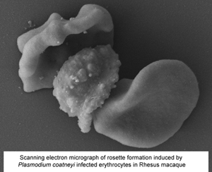
Plasmodium coatneyi has been proposed as an animal model for human Plasmodium falciparum malaria as it appears to replicate many aspects of pathogenesis and clinical symptomology. As part of the ongoing evaluation of the rhesus macaque model of severe malaria, a detailed ultrastructural analysis of the interaction between the parasite and both the host erythrocytes and the microvasculature was undertaken. Tissue (brain, heart and kidney) from splenectomized rhesus macaques and blood from spleen-intact animals infected with P. coatneyi were examined by electron microscopy. In all three tissues, similar interactions (sequestration) between infected red blood cells (iRBC) and blood vessels were observed with evidence of rosette and auto-agglutinate formation. The iRBCs possessed caveolae similar to P. vivax and knob-like structures similar to P. falciparum. However, the knobs often appeared incompletely formed in the splenectomized animals in contrast to the intact knobs exhibited by spleen intact animals. Plasmodium coatneyi infection in the monkey replicates many of the ultrastructural features particularly associated with P. falciparum in humans and as such supports its use as a suitable animal model. However, the possible effect on host–parasite interactions and the pathogenesis of disease due to the use of splenectomized animals needs to be taken into consideration.

Type
Research Article
Creative Commons
This is a work of the US Government and is not subject to copyright protection within the United States. Published by Cambridge University Press.
Copyright
Copyright © United States Army - Armed Forces Research Institute of Medical Sciences (AFRIMS), 2021

Access options

Get access to the full version of this content by using one of the access options below. (Log in options will check for institutional or personal access. Content may require purchase if you do not have access.)

References

Aikawa, M (1988 a) Human cerebral malaria. American Journal of Tropical Medicine and Hygiene 39, 110.CrossRefGoogle ScholarPubMed
Aikawa, M (1988 b) Morphological changes in erythrocytes induced by malarial parasites. Biology of the Cell 64, 173181.CrossRefGoogle ScholarPubMed
Aikawa, M, Miller, LH and Rabbege, J (1975) Caveola-vesicle complexes in the plasmalemma of erythrocytes infected by Plasmodium vivax and P. cynomolgi. Unique structures related to Schüffner's dots. American Journal of Pathology 79, 285300.Google ScholarPubMed
Aikawa, M, Rabbege, JR, Udeinya, I and Miller, LH (1983) Electron microscopy of knobs in Plasmodium falciparum-infected erythrocytes. Journal of Parasitology 69, 435437.CrossRefGoogle ScholarPubMed
Aikawa, M, Udeinya, IJ, Rabbege, J, Dayan, M, Leech, JH, Howard, RJ and Miller, LH (1985) Structural alteration of the membrane of erythrocytes infected with Plasmodium falciparum. Journal of Protozoology 32, 424429.CrossRefGoogle ScholarPubMed
Aikawa, M, Brown, AE, Smith, CD, Tegoshi, T, Howard, RJ, Hasler, TH, Ito, Y, Collins, WE and Webster, HK (1992) Plasmodium coatneyi-infected rhesus monkeys: a primate model for human cerebral malaria. Instituto Oswaldo Cruz 87(Suppl. 3), 443447.10.1590/S0074-02761992000700074CrossRefGoogle ScholarPubMed
Akinyi, S, Hanssen, E, Meyer, EV, Jiang, J, Korir, CC, Singh, B, Lapp, S, Barnwell, JW, Tilley, L and Galinski, MR (2012) A 95 kDa protein of Plasmodium vivax and P. cynomolgi visualized by three-dimensional tomography in the caveola-vesicle complexes (Schuffner's dots) of infected erythrocytes is a member of the PHIST family. Molecular Microbiology 84, 816831.CrossRefGoogle ScholarPubMed
Atkinson, CT and Aikawa, M (1990) Ultrastructure of malaria-infected erythrocytes. Blood Cells 16, 351368.Google ScholarPubMed
Bannister, LH, Hopkins, JM, Fowler, RE, Krishna, S and Mitchell, GH (2000) A brief illustrated guide to the ultrastructure of Plasmodium falciparum asexual blood stages. Parasitology Today 16, 427433.CrossRefGoogle Scholar
Barnwell, JW, Ingravallo, P, Galinski, MR, Matsumoto, Y and Aikawa, M (1990) Plasmodium vivax: malarial proteins associated with the membrane-bound caveola-vesicle complexes and cytoplasmic cleft structures of infected erythrocytes. Experimental Parasitology 70, 8599.CrossRefGoogle ScholarPubMed
Carlson, J (1993) Erythrocyte rosetting in Plasmodium falciparum malaria-with special reference to the pathogenesis of cerebral malaria. Scandinavian Journal of Infectious Diseases Supplemental 86, 179.Google Scholar
Carlson, J, Helmby, H, Hill, AV, Brewster, D, Greenwood, BM and Wahlgren, M (1990) Human cerebral malaria: association with erythrocyte rosetting and lack of anti-rosetting antibodies. Lancet (London, England) 336, 14571460.CrossRefGoogle ScholarPubMed
Cooke, BM, Mohandas, N and Coppel, RL (2004) Malaria and the red blood cell membrane. Hematology (Amsterdam, Netherlands) 41, 173188.Google ScholarPubMed
David, PH, Handunnetti, SM, Leech, JH, Gamage, P and Mendis, KN (1988) Rosetting: a new cytoadherence property of malaria-infected erythrocytes. American Journal of Tropical Medicine and Hygiene 38, 289297.10.4269/ajtmh.1988.38.289CrossRefGoogle ScholarPubMed
Desowitz, RS, Miller, LH, Buchanan, RD, Yuthasastrkosol, V and Permpanich, B (1967) Comparative studies on the pathology and host physiology of malarias: I. Plasmodium coatneyi. Annals of Tropical Medicine and Parasitology 61, 365374.10.1080/00034983.1967.11686501CrossRefGoogle ScholarPubMed
Desowitz, RS, Miller, LH, Buchanan, RD and Permpanich, B (1969) The sites of deep vascular schizogony in Plasmodium coatneyi malaria. Transactions of The Royal Society of Tropical Medicine and Hygiene 63, 198202.CrossRefGoogle ScholarPubMed
Dörpinghaus, M, Fürstenwerth, F, Roth, LK, Bouws, P, Rakotonirinalalao, M, Jordan, V, Sauer, M, Rehn, T, Pansegrau, E, Höhn, K, Mesén-Ramírez, P, Bachmann, A, Lorenzen, S, Roeder, T, Metwally, NG and Bruchhaus, I (2020) Stringent selection of knobby Plasmodium falciparum-infected erythrocytes during cytoadhesion at febrile temperature. Microorganisms 8, 174.CrossRefGoogle ScholarPubMed
Escalante, AA, Barrio, E and Ayala, FJ (1995) Evolutionary origin of human and primate malarias: evidence from the circumsporozoite protein gene. Molecular Biology and Evolution 12, 616626.Google ScholarPubMed
Eyles, DE, Fong, YL, Warren, M, Guinn, E, Sandosham, AA and Wharton, R (1962) Plasmodium coatneyi, a new species of primate malaria from Malaya. American Journal of Tropical Medicine and Hygiene 11, 597604.10.4269/ajtmh.1962.11.597CrossRefGoogle Scholar
Eyles, DE, Dunn, FL, Warren, M and Guinn, E (1963) Plasmodium coatneyi from the Philippines. Journal of Parasitology 49, 1038.CrossRefGoogle ScholarPubMed
Eyles, DE, Fong, YL, Warren, M, Guinn, EG, Sandosham, AA and Wharton, RH (1971) Plasmodium coatneyi. In Coatney, GR, Collins, WE, Warren, M and Contacos, PG (eds), The Primate Malarias. Washington, DC, USA: Government Printing Office, pp. 289299.Google Scholar
Fernandez-Becerra, C, Bernabeu, M, Castellanos, A, Bruna, R, Correa, BR, Obadia, T, Ramirez, M, Rui, E, Hentzschel, F, López-Montañés, M, Ayllon-Hermida, A, Martin-Jaular, L, Elizalde-Torrent, A, Siba, P, Vêncio, RZ, Arevalo-Herrera, M, Herrera, S, Alonso, PL, Mueller, I and del Portillo, HA (2020) Plasmodium vivax spleen-dependent genes encode antigens associated with cytoadhesion and clinical protection. Proceedings of the National Academy of Sciences USA 117, 1305613065.CrossRefGoogle ScholarPubMed
Fernandez, V and Wahlgren, M (2002) Rosetting and autoagglutination in Plasmodium falciparum. Chemical Immunology 80, 163187.Google ScholarPubMed
Handunnetti, SM, David, PH, Perera, KL and Mendis, KN (1989) Uninfected erythrocytes form “rosettes” around Plasmodium falciparum infected erythrocytes. American Journal of Tropical Medicine and Hygiene 40, 115118.CrossRefGoogle ScholarPubMed
Hasler, T, Handunnetti, SM, Aguiar, JC, van Schravendijk, MR, Greenwood, BM, Lalliger, G, Cegielski, P and Howard, RJ (1990) In vitro rosetting, cytoadherence, and microagglutination properties of Plasmodium falciparum-infected erythrocytes from Gambian and Tanzanian patients. Blood 76, 18451852.CrossRefGoogle ScholarPubMed
Hayakawa, T, Culleton, R, Otani, H, Horii, T and Tanabe, K (2008) Big bang in the evolution of extant malaria parasites. Molecular Biology and Evolution 25, 22332239.CrossRefGoogle Scholar
Ho, M, Davis, TM, Silamut, K, Bunnag, D and White, NJ (1991) Rosette formation of Plasmodium falciparum-infected erythrocytes from patients with acute malaria. Infection and Immunity 59, 21352139.10.1128/iai.59.6.2135-2139.1991CrossRefGoogle ScholarPubMed
Horrocks, P, Pinches, RA, Chakravorty, SJ, Papakrivos, J, Christodoulou, Z, Kyes, SE, Urban, BC, Ferguson, DJP and Newbold, CI (2005) PfEMP1 expression is reduced on the surface of knobless Plasmodium falciparum infected erythrocytes. Journal of Cell Science 118(Pt 11), 25072518.10.1242/jcs.02381CrossRefGoogle ScholarPubMed
Kawai, S, Aikawa, M, Kano, S and Suzuki, M (1993) A primate model for severe human malaria with cerebral involvement: Plasmodium coatneyi-infected Macaca fuscata. American Journal of Tropical Medicine and Hygiene 48, 630636.CrossRefGoogle ScholarPubMed
Kawai, S, Kano, S and Suzuki, M (1995) Rosette formation by Plasmodium coatneyi-infected erythrocytes of the Japanese macaque (Macaca fuscata). American Journal of Tropical Medicine and Hygiene 53, 295299.CrossRefGoogle Scholar
Kawai, S, Aikawa, M, Suzuki, M and Matsuda, H (1998) A nonhuman primate model for severe human malaria: Plasmodium coatneyi-infected Japanese macaque (Macaca fuscata). The Tokai Journal of Experimental and Clinical Medicine 23, 101102.Google Scholar
Kawai, S, Matsumoto, J, Aikawa, M and Matsuda, H (2003) Increased plasma levels of soluble intercellular adhesion molecule-1 (sICAM-1) and soluble vascular cell molecule-1 (sVCAM-1) associated with disease severity in a primate model for severe human malaria: Plasmodium coatneyi-infected Japanese macaques (Macaca fuscata). Journal of Veterinary Medical Science 65, 629631.CrossRefGoogle Scholar
Kho, S, Qotrunnada, L, Leonardo, L, Andries, B, Wardani, PAI, Fricot, A, Henry, B, Hardy, D, Margyaningsih, NI, Apriyanti, D, Puspitasari, AM, Prayoga, P, Trianty, L, Kenangalem, E, Chretien, F, Brousse, V, Safeukui, I, del Portillo, HA, Fernandez-Becerra, C, Meibalan, E, Marti, M, Price, RN, Woodberry, T, Ndour, PA, Russell, BM, Yeo, TW, Minigo, G, Noviyanti, R, Poespoprodjo, JR, Siregar, NC, Buffet, PA and Anstey, NM (2021) Evaluation of splenic accumulation and colocalization of immature reticulocytes and Plasmodium vivax in asymptomatic malaria: a prospective human splenectomy study. PLoS Medicine 18, e1003632.CrossRefGoogle ScholarPubMed
Kilejian, A, Abati, A and Trager, W (1977) Plasmodium falciparum and Plasmodium coatneyi: immunogenicity of “knob-like protrusions” on infected erythrocyte membranes. Experimental Parasitology 42, 157164.CrossRefGoogle ScholarPubMed
Langreth, SG, Reese, RT, Motyl, MR and Trager, W (1979) Plasmodium falciparum: loss of knobs on the infected erythrocyte surface after long-term cultivation. Experimental Parasitology 48, 213219.10.1016/0014-4894(79)90101-2CrossRefGoogle ScholarPubMed
Leclerc, MC, Hugot, JP, Durand, P and Renaud, F (2004) Evolutionary relationships between 15 Plasmodium species from new and old-world primates (including humans): an 18S rDNA cladistic analysis. Parasitology 129(Pt 6), 677684.CrossRefGoogle ScholarPubMed
Lee, W-C, Russell, B, Lau, Y-L, Fong, M-Y, Chu, C, Sriprawat, K, Suwanarusk, R, Nosten, F and Renia, L (2013) Giemsa-stained wet mount based method for reticulocyte quantification: a viable alternative in resource-limited or malaria-endemic settings. PLoS ONE 8, e60303.CrossRefGoogle ScholarPubMed
Lee, WC, Malleret, B, Lau, Y-L, Mauduit, M, Fong, M-Y, Cho, JS, Suwanarusk, R, Zhang, R, Albrecht, L, Costa, FTM, Preiser, P, McGready, R, Renia, L, Nosten, F and Russell, B (2014) Glycophorin C (CD236R) mediates vivax malaria parasite rosetting to normocytes [published correction appears in Blood. 2015; 126, 2765]. Blood 123, e100e109.CrossRefGoogle Scholar
Liu, B, Blanch, AJ, Namvar, A, Carmo, O, Tiash, S, Andrew, D, Hanssen, E, Rajagopal, V, Dixon, MWA and Tilley, L (2019) Multimodal analysis of Plasmodium knowlesi-infected erythrocytes reveals large invaginations, swelling of the host cell, and rheological defects. Cell Microbiology 21, e13005.CrossRefGoogle ScholarPubMed
Lombardini, ED, Gettayacamin, M, Turner, GDH and Brown, AE (2015) A review of Plasmodium coatneyi-macaque models of severe and cerebral malaria. Veterinary Pathology 52, 9981011.CrossRefGoogle Scholar
Lombardini, ED, Turner, GDH, Brown, AE, Inamnuay, L, Kaewamatawong, T, Sunyakumthorn, P and Ferguson, DJP (2021) Systemic ultrastructural tissue pathology of Plasmodium coatneyi infection in rhesus macaques. Veterinary Pathology (accepted for publication).Google Scholar
MacPherson, GG, Warrell, MJ, White, NJ, Looareesuwan, S and Warrell, DA (1985) Human cerebral malaria. A quantitative ultrastructural analysis of parasitized erythrocyte sequestration. American Journal of Pathology 119, 385401.Google ScholarPubMed
Maeno, Y, Brown, AE, Smith, CD, Tegoshi, T, Toyoshima, T, Ockenhouse, C, Corcoran, K, Ngampochjana, M, Kyle, D and Webster, H (1993) A non-human primate model for cerebral malaria: effects of artesunate (qinghaosu derivative) on rhesus monkeys experimentally infected with Plasmodium coatneyi. American Journal of Tropical Medicine and Hygiene 49, 726734.CrossRefGoogle Scholar
Malleret, B, Xu, F, Mohandas, N, Suwanarusk, R, Chu, C, Leite, JA, Low, K, Turner, C, Sriprawat, K, Zhang, R, Bertrand, O, Colin, Y, Costa, FTM, Ong, CN, Ng, ML, Lim, CT, Nosten, F, Renia, L and Russell, B (2013) Significant biochemical, biophysical and metabolic diversity in circulating human cord blood reticulocytes. PLoS One 8, e76062.CrossRefGoogle ScholarPubMed
Moreno, A, Cabrera-Mora, M, Garcia, A, Orkin, J, Strobert, E, Barnwell, JW and Galinski, MR (2013) Plasmodium coatneyi in rhesus macaques replicates the multisystemic dysfunction of severe malaria in humans. Infection and Immunity 81, 18891904.CrossRefGoogle ScholarPubMed
Mundwiler-Pachlatko, E and Beck, HP (2013) Maurer's clefts, the enigma of Plasmodium falciparum. Proceedings of the National Academy of Sciences USA 110, 1998719994.CrossRefGoogle ScholarPubMed
Nguansangiam, S, Day, NP, Hien, TT, Mai, NTH, Chaisri, U, Riganti, M, Dondorp, AM, Lee, SJ, Phu, NH, Turner, GDH, White, NJ, Ferguson, DJP and Pongponratn, E (2007) A quantitative ultrastructural study of renal pathology in fatal Plasmodium falciparum malaria. Tropical Medicine and International Health 12, 10371050.CrossRefGoogle ScholarPubMed
Pongponratn, E, Riganti, M, Punpoowong, B and Aikawa, M (1991) Microvascular sequestration of parasitized erythrocytes in human falciparum malaria: a pathological study. American Journal of Tropical Medicine and Hygiene 44, 168175.CrossRefGoogle ScholarPubMed
Pongponratn, E, Viriyavejakul, P, Wilairatana, P, Ferguson, DJP, Chaisri, U, Turner, GDH and Looareesuwan, S (2000) Absence of knobs on parasitized red blood cells in a splenectomized patient in fatal falciparum malaria. Southeast Asian Journal of Tropical Medicine and Public Health 31, 829835.Google Scholar
Pongponratn, E, Turner, GDH, Day, NP, Phu, NH, Simpson, JA, Stepniewska, K, Mai, NTH, Viriyavejakul, P, Looareesuwan, S, Hien, TT, Ferguson, DJP and White, NJ (2003) An ultrastructural study of the brain in fatal Plasmodium falciparum malaria. American Journal of Tropical Medicine and Hygiene 69, 345359.CrossRefGoogle ScholarPubMed
Prommano, O, Chaisri, U, Turner, GDH, Wilairatana, P, Ferguson, DJP, Viriyavejakul, P, White, NJ and Pongponratn, E (2005) A quantitative ultrastructural study of the liver and the spleen in fatal falciparum malaria. Southeast Asian Journal of Tropical Medicine and Public Health 36, 13591370.Google ScholarPubMed
Quadt, KA, Barfod, L, Andersen, D, Bruun, J, Gyan, B, Hassenkam, T, Ofori, MF and Hviid, L (2012) The density of knobs on Plasmodium falciparum-infected erythrocytes depends on developmental age and varies among isolates. PLoS One 7, e45658.CrossRefGoogle ScholarPubMed
Roberts, DJ, Pain, A, Kai, O, Kortok, M and Marsh, K (2000) Autoagglutination of malaria-infected red blood cells and malaria severity. Lancet (London, England) 355, 14271428.CrossRefGoogle ScholarPubMed
Sein, KK, Brown, AE, Maeno, Y, Smith, CD, Corcoran, KD, Hansukjariya, P, Webster, HK and Aikawa, M (1993) Sequestration pattern of parasitized erythrocytes in cerebrum, mid-brain and cerebellum of Plasmodium coatneyi-infected rhesus monkeys (Macaca mulatta). American Journal of Tropical Medicine and Hygiene 49, 513519.10.4269/ajtmh.1993.49.513CrossRefGoogle Scholar
Smith, CD, Brown, AE, Nakazawa, S, Fujioka, H and Aikawa, M (1996) Multi-organ erythrocyte sequestration and ligand expression in rhesus monkeys infected with Plasmodium coatneyi malaria. American Journal of Tropical Medicine and Hygiene 55, 379383.CrossRefGoogle ScholarPubMed
Subramani, R, Quadt, K, Jeppesen, AE, Hempel, C, Petersen, JEV, Hassenkam, T, Hviid, L and Barfod, L (2015) Plasmodium falciparum-infected erythrocyte knob density is linked to the PfEMP1 variant expressed. MBio 6, e01456-15.CrossRefGoogle Scholar
Tegoshi, T, Udomsangpetch, R, Brown, AE, Nakazawa, S, Webster, HK and Aikawa, M (1993) Ultrastructure of rosette formation by Plasmodium coatneyi-infected erythrocytes of rhesus. Parasitology Research 79, 611613.10.1007/BF00932248CrossRefGoogle ScholarPubMed
Tilly, AK, Thiede, J, Metwally, N, Lubiana, P, Bachmann, A, Roeder, T, Rockliffe, N, Lorenzen, S, Tannich, E, Gutsmann, T and Bruchhaus, I (2015) Type of in vitro cultivation influences cytoadhesion, knob structure, protein localization and transcriptome profile of Plasmodium falciparum. Scientific Reports 5, 16766.CrossRefGoogle ScholarPubMed
Udagama, PV, Atkinson, CT, Peiris, JS, David, PH, Mendis, KN and Aikawa, M (1988) Immunoelectron microscopy of Schuffner's dots in Plasmodium vivax-infected human erythrocytes. American Journal of Pathology 131, 4852.Google ScholarPubMed
Udomsangpetch, R, Wåhlin, B, Carlson, J, Berzins, K, Torri, M, Aikawa, M, Perlmann, P and Wahlgren, M (1989) Plasmodium falciparum-infected erythrocytes form spontaneous erythrocyte rosettes. Journal of Experimental Medicine 169, 18351840.CrossRefGoogle ScholarPubMed
Udomsangpetch, R, Brown, AE, Smith, CD and Webster, HK (1991) Rosette formation by Plasmodium coatneyi-infected red blood cells. American Journal of Tropical Medicine and Hygiene 44, 399401.CrossRefGoogle ScholarPubMed
Wahlgren, M, Carlson, J, Udomsangpetch, R and Perlmann, P (1989) Why do Plasmodium falciparum-infected erythrocytes form spontaneous erythrocyte rosettes? Parasitology Today 5, 183185.CrossRefGoogle Scholar
Watermeyer, JM, Hale, VL, Hackett, F, Clare, DK, Cutts, EE, Vakonakis, I, Fleck, RA, Blackman, MJ and Saibil, HR (2016) A spiral scaffold underlies cytoadherent knobs in Plasmodium falciparum-infected erythrocytes. Blood 127, 343351.CrossRefGoogle ScholarPubMed