Hostname: page-component-745bb68f8f-b95js Total loading time: 0 Render date: 2025-01-14T10:21:00.106Z Has data issue: false hasContentIssue false

Shooting for the moon: using tissue-mimetic hydrogels to gain new insight on cancer biology and screen therapeutics

Published online by Cambridge University Press:  07 September 2017

Samantha E. Holt
Affiliation:
Department of Biomedical Engineering, Texas A&M University, College Station, Texas, USA
E. Sally Ward
Affiliation:
Department of Molecular and Cellular Medicine, Texas A&M Health Science Center, College Station, Texas, USA
Raimund J. Ober
Affiliation:
Department of Biomedical Engineering, Texas A&M University, College Station, Texas, USA Department of Molecular and Cellular Medicine, Texas A&M Health Science Center, College Station, Texas, USA
Daniel L. Alge*
Affiliation:
Department of Biomedical Engineering, Texas A&M University, College Station, Texas, USA Department of Materials Science and Engineering, Texas A&M University, College Station, Texas, USA
*
Address all correspondence to D. L. Alge at dalge@tamu.edu
Get access

Abstract

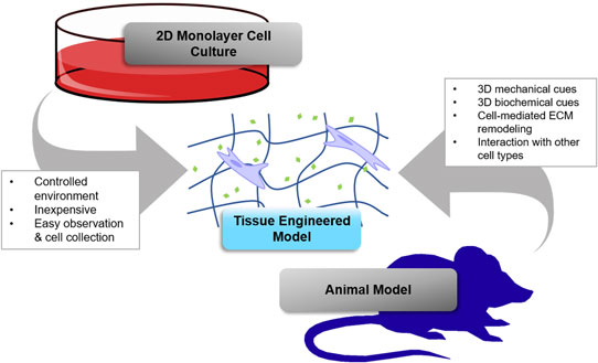
Tissue engineering holds great promise for advancing cancer research and achieving the goals of the Cancer Moonshot by providing better models for basic research and testing novel therapeutics. This paper focuses on the use of hydrogel biomaterials due to their unique ability to entrap cells in three-dimensional (3D) matrix that mimics tissues and can be programmed with physical and chemical cues to recreate key aspects of tumor microenvironments. The chemistry of some commonly used hydrogel platforms is discussed, and important examples of their use in tissue engineering 3D cancer models are highlighted. Challenges and opportunities for future research are also discussed.

Type
Biomaterials for 3D Cell Biology Prospective Articles
Copyright
Copyright © Materials Research Society 2017 

Access options

Get access to the full version of this content by using one of the access options below. (Log in options will check for institutional or personal access. Content may require purchase if you do not have access.)

References

1.Hanahan, D. and Weinberg, R.A.: Hallmarks of cancer: the next generation. Cell 144, 646 (2011).Google Scholar
2.Schuessler, T.K., Chan, X.Y., Chen, H.J., Ji, K., Park, K.M., Roshan-Ghias, A., Sethi, P., Thakur, A., Tian, X., Villasante, A., Zervantonakis, I.K., Moore, N.M., Nagahara, L.A., and Kuhn, N.Z.: Biomimetic tissue-engineered systems for advancing cancer research: NCI strategic workshop report. Cancer Res. 74, 5359 (2014).Google Scholar
3.Hahn, W.C., Stewart, S.A., Brooks, M.W., York, S.G., Eaton, E., Kurachi, A., Beijersbergen, R.L., Knoll, J.H.M., Meyerson, M., and Weinberg, R.A.: Inhibition of telomerase limits the growth of human cancer cells. Nat. Med. 5, 1164 (1999).Google Scholar
4.Leight, J.L., Wozniak, M.A., Chen, S., Lynch, M.L., and Chen, C.S.: Matrix rigidity regulates a switch between TGF-β1-induced apoptosis and epithelial-mesenchymal transition. Mol. Biol. Cell 23, 781 (2012).Google Scholar
5.Heyer, J., Kwong, L.N., Lowe, S.W., and Chin, L.: Non-germline genetically engineered mouse models for translational cancer research. Nat. Rev. Cancer 10, 470 (2010).Google Scholar
6.Sharpless, N.E. and DePinho, R.A.: The mighty mouse: genetically engineered mouse models in cancer drug development. Nat. Rev. Drug Discov. 5, 741 (2006).Google Scholar
7.Rangarajan, A. and Weinberg, R.A.: Comparative biology of mouse versus human cells: modelling human cancer in mice. Nat. Rev. Cancer 3, 952 (2003).Google Scholar
8.Mak, I.W.Y., Evaniew, N., and Ghert, M.: Lost in translation: animal models and clinical trials in cancer treatment. Am. J. Transl. Res. 6, 114 (2014).Google Scholar
9.Petersen, O.W., Rønnov-Jessen, L., Howlett, A.R., and Bissell, M.J.: Interaction with basement membrane serves to rapidly distinguish growth and differentiation pattern of normal and malignant human breast epithelial cells. Proc. Natl. Acad. Sci. U. S. A. 89, 9064 (1992).Google Scholar
10.Debnath, J., Muthuswamy, S.K., and Brugge, J.S.: Morphogenesis and oncogenesis of MCF-10A mammary epithelial acini grown in three-dimensional basement membrane cultures. Methods 30, 256 (2003).Google Scholar
11.Albini, A., Iwamoto, I., Kleinman, H.K., Martin, G.R., Aaronson, S.A., Kozlowski, J.M., and McEwan, R.N.: A rapid in vitro assay for quantitating the invasive potential of tumor cells. Cancer Res. 47, 3239 (1987).Google Scholar
12.Hughes, C.S., Postovit, L.M., and Lajoie, G.A.: Matrigel: a complex protein mixture required for optimal growth of cell culture. Proteomics 10, 1886 (2010).Google Scholar
13.Hoffman, A.S.: Hydrogels for biomedical applications. Adv. Drug Deliv. Rev. 64, 18 (2012).Google Scholar
14.Magin, C.M., Alge, D.L., and Anseth, K.S.: Bio-inspired 3D microenvironments: a new dimension in tissue engineering. Biomed. Mater. 11, 22001 (2016).Google Scholar
15.Azagarsamy, M.A. and Anseth, K.S.: Bioorthogonal click chemistry: an indispensable tool to create multifaceted cell culture scaffolds. ACS Macro Lett. 2, 5 (2013).Google Scholar
16.Nguyen, K.T. and West, J.L.: Photopolymerizable hydrogels for tissue engineering applications. Biomaterials 23, 4307 (2002).Google Scholar
17.Lee, K.Y. and Mooney, D.J.: Hydrogels for tissue engineering. Chem. Rev. 101, 1869 (2001).Google Scholar
18.Fairbanks, B.D., Schwartz, M.P., Bowman, C.N., and Anseth, K.S.: Photoinitiated polymerization of PEG-diacrylate with lithium phenyl-2,4,6-trimethylbenzoylphosphinate: polymerization rate and cytocompatibility. Biomaterials 30, 6702 (2009).Google Scholar
19.Nimmo, C.M. and Shoichet, M.S.: Regenerative biomaterials that “click”: simple, aqueous-based protocols for hydrogel synthesis, surface immobilization, and 3D patterning. Bioconjug. Chem. 22, 2199 (2011).Google Scholar
20.Lin, C.-C., Raza, A., and Shih, H.: PEG hydrogels formed by thiol-ene photo-click chemistry and their effect on the formation and recovery of insulin-secreting cell spheroids. Biomaterials 32, 9685 (2011).Google Scholar
21.Shubin, A.D., Felong, T.J., Graunke, D., Ovitt, C.E., and Benoit, D.S.W.: Development of poly(ethylene glycol) hydrogels for salivary gland tissue engineering applications. Tissue Eng. A 21, 1733 (2015).Google Scholar
22.Parenteau-Bareil, R., Gauvin, R., and Berthod, F.: Collagen-based biomaterials for tissue engineering applications. Materials (Basel) 3, 1863 (2010).Google Scholar
23.Loessner, D., Meinert, C., Kaemmerer, E., Martine, L.C., Yue, K., Levett, P.A., Klein, T.J., Melchels, F.P.W., Khademhosseini, A., and Hutmacher, D.W.: Functionalization, preparation and use of cell-laden gelatin methacryloyl-based hydrogels as modular tissue culture platforms. Nat. Protoc. 11, 727 (2016).Google Scholar
24.Xu, X., Jha, A.K., Harrington, D.A., Farach-Carson, M.C., and Jia, X.: Hyaluronic acid-based hydrogels: from a natural polysaccharide to complex networks. Soft Matter 8, 3280 (2012).Google Scholar
25.Augst, A.D., Kong, H.J., and Mooney, D.J.: Alginate hydrogels as biomaterials. Macromol. Biosci. 6, 623 (2006).Google Scholar
26.Jeon, O., Bouhadir, K.H., Mansour, J.M., and Alsberg, E.: Photocrosslinked alginate hydrogels with tunable biodegradation rates and mechanical properties. Biomaterials 30, 2724 (2009).Google Scholar
27.Zhu, J.: Bioactive modification of poly(ethylene glycol) hydrogels for tissue engineering. Biomaterials 31, 4639 (2010).Google Scholar
28.Lutolf, M.P., Lauer-Fields, J.L., Schmoekel, H.G., Metters, T., Weber, F.E., Fields, G.B., and Hubbell, J.: Synthetic matrix metalloproteinase-sensitive hydrogels for the conduction of tissue regeneration: engineering cell-invasion characteristics. Proc. Natl. Acad. Sci. U. S. A. 100, 5413 (2003).Google Scholar
29.Phelps, E.A., Enemchukwu, N.O., Fiore, V.F., Sy, J.C., Murthy, N., Sulchek, T.A., Barker, T.H., and Garcia, A.J.: Maleimide cross-linked bioactive PEG hydrogel exhibits improved reaction kinetics and cross-linking for cell encapsulation and in situ delivery. Adv. Mater. 24, 64 (2012).Google Scholar
30.Rydholm, A.E., Anseth, K.S., and Bowman, C.N.: Effects of neighboring sulfides and pH on ester hydrolysis in thiol-acrylate photopolymers. Acta Biomater. 3, 449 (2007).Google Scholar
31.Fairbanks, B.D., Schwartz, M.P., Halevi, A.E., Nuttelman, C.R., Bowman, C.N., and Anseth, K.S.: A versatile synthetic extracellular matrix mimic via Thiol-Norbornene photopolymerization. Adv. Mater. 21, 5005 (2009).Google Scholar
32.Gill, B.J., Gibbons, D.L., Roudsari, L.C., Saik, J.E., Rizvi, Z.H., Roybal, J.D., Kurie, J.M., and West, J.L.: A synthetic matrix with independently tunable biochemistry and mechanical properties to study epithelial morphogenesis and EMT in a lung adenocarcinoma model. Cancer Res. 72, 6013 (2012).Google Scholar
33.Roudsari, L.C., Jeffs, S.E., Witt, A.S., Gill, B.J., and West, J.L.: A 3D poly(ethylene glycol)-based tumor angiogenesis model to study the influence of vascular cells on lung tumor cell behavior. Sci. Rep. 6, 32726 (2016).Google Scholar
34.Ki, C.S., Lin, T.-Y., Korc, M., and Lin, C.-C.: Thiol-ene hydrogels as desmoplasia-mimetic matrices for modeling pancreatic cancer cell growth, invasion, and drug resistance. Biomaterials 35, 9668 (2014).Google Scholar
35.Liang, Y., Jeong, J., DeVolder, R.J., Cha, C., Wang, F., Tong, Y.W., and Kong, H.: A cell-instructive hydrogel to regulate malignancy of 3D tumor spheroids with matrix rigidity. Biomaterials 32, 9308 (2011).Google Scholar
36.Xu, X., Liu, C., Liu, Y., Li, N., Guo, X., Wang, S., Sun, G., Wang, W., and Ma, X.: Encapsulated human hepatocellular carcinoma cells by alginate gel beads as an in vitro metastasis model. Exp. Cell Res. 319, 2135 (2013).Google Scholar
37.Lin, T.-Y., Ki, C.S., and Lin, C.-C.: Manipulating hepatocellular carcinoma cell fate in orthogonally cross-linked hydrogels. Biomaterials 35, 6898 (2014).Google Scholar
38.Fischbach, C., Kong, H.J., Hsiong, S.X., Evangelista, M.B., Yuen, W., and Mooney, D.J.: Cancer cell angiogenic capability is regulated by 3D culture and integrin engagement. Proc. Natl. Acad. Sci. U. S. A. 106, 399 (2009).Google Scholar
39.Sieh, S., Taubenberger, A.V., Rizzi, S.C., Sadowski, M., Lehman, M.L., Rockstroh, A., An, J., Clements, J.A., Nelson, C.C., and Hutmacher, D.W.: Phenotypic characterization of prostate cancer LNCaP cells cultured within a bioengineered microenvironment. PLoS ONE 7, e40217 (2012).Google Scholar
40.Xu, X., Gurski, L.A., Zhang, C., Harrington, D.A., Farach-Carson, M.C., and Jia, X.: Recreating the tumor microenvironment in a bilayer, hyaluronic acid hydrogel construct for the growth of prostate cancer spheroids. Biomaterials 33, 9049 (2012).Google Scholar
41.Loessner, D., Stok, K.S., Lutolf, M.P., Hutmacher, D.W., Clements, J.A., and Rizzi, S.C.: Bioengineered 3D platform to explore cell-ECM interactions and drug resistance of epithelial ovarian cancer cells. Biomaterials 31, 8494 (2010).Google Scholar
42.Yang, Z. and Zhao, X.: A 3D model of ovarian cancer cell lines on peptide nanofiber scaffold to explore the cell-scaffold interaction and chemotherapeutic resistance of anticancer drugs. Int. J. Nanomed. 5, 303 (2011).Google Scholar
43.Kaemmerer, E., Melchels, F.P.W., Holzapfel, B.M., Meckel, T., Hutmacher, D.W., and Loessner, D.: Gelatine methacrylamide-based hydrogels: an alternative three-dimensional cancer cell culture system. Acta Biomater. 10, 2551 (2014).Google Scholar
44.Holmes, D.: The cancer that rises with the Sun. Nature 515, S110 (2014).Google Scholar
45.Glazer, A.M., Winkelmann, R.R., Farberg, A.S., and Rigel, D.S.: Analysis of trends in US melanoma incidence and mortality. JAMA Dermatol. 153, 225 (2017).Google Scholar
46.Reed, K.B., Brewer, J.D., Lohse, C.M., Bringe, K.E., Pruitt, C.N., and Gibson, L.E.: Increasing incidence of melanoma among young adults: an epidemiological study in Olmsted County, Minnesota. Mayo Clin. Proc. 87, 328 (2012).Google Scholar
47.Tokuda, E.Y., Leight, J.L., and Anseth, K.S.: Modulation of matrix elasticity with PEG hydrogels to study melanoma drug responsiveness. Biomaterials 35, 4310 (2014).Google Scholar
48.Singh, S.P., Schwartz, M.P., Tokuda, E.Y., Luo, Y., Rogers, R.E., Fujita, M., Ahn, N.G., and Anseth, K.S.: A synthetic modular approach for modeling the role of the 3D microenvironment in tumor progression. Sci. Rep. 5, 1 (2015).Google Scholar
49.Tokuda, E.Y., Jones, C.E., and Anseth, K.S.: PEG-peptide hydrogels reveal differential effects of matrix microenvironmental cues on melanoma drug sensitivity. Integr. Biol. 9, 76 (2017).Google Scholar
50.Kalluri, R. and Zeisberg, M.: Fibroblasts in cancer. Nat. Rev. Cancer 6, 392 (2006).Google Scholar
51.Leight, J.L., Tokuda, E.Y., Jones, C.E., Lin, A.J., and Anseth, K.S.: Multifunctional bioscaffolds for 3D culture of melanoma cells reveal increased MMP activity and migration with BRAF kinase inhibition. Proc. Natl. Acad. Sci. U. S. A. 112, 5366 (2015).Google Scholar
52.Sosman, J.A., Kim, K.B., Schuchter, L., Gonzalez, R., Pavlick, A.C., Weber, J.S., McArthur, G.A., Hutson, T.E., Moschos, S.J., Flaherty, K.T., Hersey, P., Kefford, R., Lawrence, D., Puzanov, I., Lewis, K.D., Amaravadi, R.K., Chmielowski, B., Lawrence, H.J., Shyr, Y., Ye, F., Li, J., Nolop, K.B., Lee, R.J., Joe, A.K., and Ribas, A.: Survival in BRAF V600-mutant advanced melanoma treated with vemurafenib. N. Engl. J. Med. 366, 707 (2012).Google Scholar
53.Thakkar, J.P., Dolecek, T.A., Horbinski, C., Ostrom, Q.T., Lightner, D.D., Barnholtz-Sloan, J.S., and Villano, J.L.: Epidemiologic and molecular prognostic review of glioblastoma. Cancer Epidemiol. Biomark. Prev. 23, 1985 (2014).Google Scholar
54.Omuro, A.M.P., Faivre, S., and Raymond, E.: Lessons learned in the development of targeted therapy for malignant gliomas. Mol. Cancer Ther. 6, 1909 (2007).Google Scholar
55.Ulrich, T.A., de Juan Pardo, E.M., and Kumar, S.: The mechanical rigidity of the extracellular matrix regulates the structure, motility, and proliferation of glioma cells. Cancer Res. 69, 4167 (2009).Google Scholar
56.Pathak, A. and Kumar, S.: Independent regulation of tumor cell migration by matrix stiffness and confinement. Proc. Natl. Acad. Sci. U. S. A. 109, 10334 (2012).Google Scholar
57.Verbridge, S.S., Choi, N.W., Zheng, Y., Brooks, D.J., Stroock, A.D., and Fischbach, C.: Oxygen-controlled three-dimensional cultures to analyze tumor angiogenesis. Tissue Eng. A 16, 2133 (2010).Google Scholar
58.Nguyen, D.T., Fan, Y., Akay, Y.M., and Akay, M.: Investigating glioblastoma angiogenesis using a 3D in vitro GelMA microwell platform. IEEE Trans. Nanobiosci. 15, 289 (2016).Google Scholar
59.Nguyen, D.T., Fan, Y., Akay, Y.M., and Akay, M.: TNP-470 reduces glioblastoma angiogenesis in three dimensional GelMA microwell platform. IEEE Trans. Nanobiosci. 15, 683 (2016).Google Scholar
60.Ananthanarayanan, B., Kim, Y., and Kumar, S.: Elucidating the mechanobiology of malignant brain tumors using a brain matrix-mimetic hyaluronic acid hydrogel platform. Biomaterials 32, 7913 (2011).Google Scholar
61.Rao, S.S., DeJesus, J., Short, A.R., Otero, J.J., Sarkar, A., and Winter, J.O.: Glioblastoma behaviors in three-dimensional collagen-hyaluronan composite hydrogels. ACS Appl. Mater. Interfaces 5, 9276 (2013).Google Scholar
62.Pedron, S., Becka, E., and Harley, B.A.C.: Regulation of glioma cell phenotype in 3D matrices by hyaluronic acid. Biomaterials 34, 7408 (2013).Google Scholar
63.Pedron, S., Becka, E., and Harley, B.A.: Spatially gradated hydrogel platform as a 3D engineered tumor microenvironment. Adv. Mater. 27, 1567 (2015).Google Scholar
64.Pedron, S. and Harley, B.A.C.: Impact of the biophysical features of a 3D gelatin microenvironment on glioblastoma malignancy. J. Biomater. Res. A 101A, 3404 (2013).Google Scholar
65.Wang, C., Tong, X., and Yang, F.: Bioengineered 3D brain tumor model to elucidate the effects of matrix stiffness on glioblastoma cell behavior using PEG-based hydrogels. Mol. Pharm. 11, 2115 (2014).Google Scholar
66.Wang, C., Tong, X., Jiang, X., and Yang, F.: Effect of matrix metalloproteinase-mediated matrix degradation on glioblastoma cell behavior in 3D PEG-based hydrogels. J. Biomed. Mater. Res. A 105A, 770 (2017).Google Scholar
67.Jiguet Jiglaire, C., Baeza-Kallee, N., Denicolaï, E., Barets, D., Metellus, P., Padovani, L., Chinot, O., Figarella-Branger, D., and Fernandez, C.: Ex vivo cultures of glioblastoma in three-dimensional hydrogel maintain the original tumor growth behavior and are suitable for preclinical drug and radiation sensitivity screening. Exp. Cell Res. 321, 99 (2014).Google Scholar
68.Siegel, R.L., Miller, K.D., and Jemal, A.: Cancer statistics, 2016. CA. Cancer J. Clin. 66, 7 (2016).Google Scholar
69.Woolston, C.: Breast cancer. Nature 527, S101 (2015).Google Scholar
70.Bleyer, A. and Welch, H.G.: Effect of three decades of screening mammography on breast-cancer incidence. N. Engl. J. Med. 367, 1998 (2012).Google Scholar
71.Pradhan, S., Hassani, I., Seeto, W.J., and Lipke, E.A.: PEG-fibrinogen hydrogels for three-dimensional breast cancer cell culture. J. Biomed. Mater. Res. A 105A, 236 (2017).Google Scholar
72.Kenny, P.A., Lee, G.Y., Myers, C.A., Neve, R.M., Semeiks, J.R., Spellman, P.T., Lorenz, K., Lee, E.H., Barcellos-Hoff, M.H., Petersen, O.W., Gray, J.W., and Bissell, M.J.: The morphologies of breast cancer cell lines in three-dimensional assays correlate with their profiles of gene expression. Mol. Oncol. 1, 84 (2007).Google Scholar
73.Pradhan, S., Clary, J.M., Seliktar, D., and Lipke, E.A.: A three-dimensional spheroidal cancer model based on PEG- fibrinogen hydrogel microspheres. Biomaterials 115, 141 (2017).Google Scholar
74.Weiss, M.S., Bernabé, B.P., Shikanov, A., Bluver, D.A., Mui, M.D., Shin, S., Broadbelt, L.J., and Shea, L.D.: The impact of adhesion peptides within hydrogels on the phenotype and signaling of normal and cancerous mammary epithelial cells. Biomaterials 33, 3548 (2012).Google Scholar
75.Taubenberger, A.V., Bray, L.J., Haller, B., Shaposhnykov, A., Binner, M., Freudenberg, U., Guck, J., and Werner, C.: 3D extracellular matrix interactions modulate tumour cell growth, invasion and angiogenesis in engineered tumour microenvironments. Acta Biomater. 36, 73 (2016).Google Scholar
76.Kloxin, A.M., Kasko, A.M., Salinas, C.N., and Anseth, K.S.: Photodegradable hydrogels for dynamic tuning of physical and chemical properties. Science 324, 59 (2009).Google Scholar
77.Wang, H., Tibbitt, M.W., Langer, S.J., Leinwand, L.A., and Anseth, K.S.: Hydrogels preserve native phenotypes of valvular fibroblasts through an elasticity-regulated PI3K/AKT pathway. Proc. Natl. Acad. Sci. U.S.A. 110, 19336 (2013).Google Scholar
78.Yang, C., Tibbitt, M.W., Basta, L., and Anseth, K.S.: Mechanical memory and dosing influence stem cell fate. Nat. Mater. 13, 645 (2014).Google Scholar
79.Stowers, R.S., Allen, S.C., Sanchez, K., Davis, C.L., Ebelt, N.D., van Den Berg, C., and Suggs, L.J.: Extracellular matrix stiffening induces a malignant phenotypic transition in breast epithelial cells. Cell. Mol. Bioeng. 10, 114 (2017).Google Scholar
80.Valdez, J., Cook, C.D., Chopko, C., Wang, A.J., Brown, A., Kumar, M., Stockdale, L., Rothenberg, D., Renggli, K., Gordon, E., Lauffenburger, D., White, F., and Griffith, L.: On-demand dissolution of modular, synthetic extracellular matrix reveals local epithelial-stromal communication networks. Biomaterials 130, 90 (2017).Google Scholar
81.Gasparian, A., Daneshian, L., Ji, H., Jabbari, E., and Shtutman, M.: Purification of high-quality RNA from synthetic polyethylene glycol-based hydrogels. Anal. Biochem. 484, 1 (2015).Google Scholar
82.Douglas, A.M., Fragkopoulos, A.A., Gaines, M.K., Lyon, L.A., Fernandez-Nieves, A., and Barker, T.H.: Dynamic assembly of ultrasoft colloidal networks enables cell invasion within restrictive fibrillar polymers. Proc. Natl. Acad. Sci. U. S. A. 114, 885 (2017).Google Scholar
83.Bray, L.J., Binner, M., Holzheu, A., Friedrichs, J., Freudenberg, U., Hutmacher, D.W., and Werner, C.: Multi-parametric hydrogels support 3D in vitro bioengineered microenvironment models of tumour angiogenesis. Biomaterials 53, 609 (2015).Google Scholar
84.Huang, S., Van Arsdall, M., Tedjarati, S., McCarty, M., Wu, W., Langley, R., and Fidler, I.J.: Contributions of stromal metalloproteinase-9 to angiogenesis and growth of human ovarian carcinoma in mice. J. Natl. Cancer Inst. 94, 1134 (2002).Google Scholar
85.Lin, E.Y., Nguyen, A. V., Russell, R.G., and Pollard, J.W.: Colony-stimulating factor 1 promotes progression of mammary tumors to malignancy. J. Exp. Med. 193, 727 (2001).Google Scholar
86.Condeelis, J. and Pollard, J.W.: Macrophages: obligate partners for tumor cell migration, invasion, and metastasis. Cell 124, 263 (2006).Google Scholar
87.Wittmann, K. and Fischbach, C.: Contextual control of adipose-derived stem cell function: implications for engineered tumor models. ACS Biomater. Sci. Eng. 3, 1483 (2017).Google Scholar
88.Heddleston, J.M., Li, Z., Lathia, J.D., Bao, S., Hjelmeland, A.B., and Rich, J.N.: Hypoxia inducible factors in cancer stem cells. Br. J. Cancer 102, 789 (2010).Google Scholar
89.Keith, B. and Simon, M.C.: Hypoxia-inducible factors, stem cells, and cancer. Cell 129, 465 (2007).Google Scholar
90.Heddleston, J.M., Li, Z., McLendon, R.E., Hjelmeland, A.B., and Rich, J.N.: The hypoxic microenvironment maintains glioblastoma stem cells and promotes reprogramming towards a cancer stem cell phenotype. Cell Cycle 8, 3274 (2009).Google Scholar
91.Conley, S.J., Gheordunescu, E., Kakarala, P., Newman, B., Korkaya, H., Heath, A.N., Clouthier, S.G., and Wicha, M.S.: Antiangiogenic agents increase breast cancer stem cells via the generation of tumor hypoxia. Proc. Natl. Acad. Sci. U. S. A. 109, 2784 (2012).Google Scholar
92.Rodenhizer, D., Gaude, E., Cojocari, D., Mahadevan, R., Frezza, C., Wouters, B.G., and McGuigan, A.P.: A three-dimensional engineered tumour for spatial snapshot analysis of cell metabolism and phenotype in hypoxic gradients. Nat. Mater. 15, 227 (2016).Google Scholar
93.Park, K.M. and Gerecht, S.: Hypoxia-inducible hydrogels. Nat. Commun. 5, article number 4075 (2014).Google Scholar
94.Lewis, D.M., Park, K.M., Tang, V., Xu, Y., Pak, K., Eisinger-Mathason, T.S.K., Simon, M.C., and Gerecht, S.: Intratumoral oxygen gradients mediate sarcoma cell invasion. Proc. Natl. Acad. Sci. U. S. A. 113, 9292 (2016).Google Scholar
95.Hartgerink, J.D., Beniash, E., and Stupp, S.I.: Peptide-amphiphile nanofibers: a versatile scaffold for the preparation of self-assembling materials. Proc. Natl. Acad. Sci. U. S. A. 99, 5133 (2002).Google Scholar
96.Collier, J.H. and Messersmith, P.B.: Self-assembling polymer-peptide conjugates: nanostructural tailoring. Adv. Mater. 16, 907 (2004).Google Scholar
97.Baker, B.M., Trappmann, B., Wang, W.Y., Sakar, M.S., Kim, I.L., Shenoy, V.B., Burdick, J.A., and Chen, C.S.: Cell-mediated fiber recruitment drives extracellular matrix mechanosensing in engineered fibrillar microenvironments. Nat. Mater. 14, 1262 (2015).Google Scholar
98.Guvendiren, M. and Burdick, J.A.: Stiffening hydrogels to probe short- and long-term cellular responses to dynamic mechanics. Nat. Commun. 3, 792 (2012).Google Scholar
99.Young, J.L. and Engler, A.J.: Hydrogels with time-dependent material properties enhance cardiomyocyte differentiation in vitro. Biomaterials 32, 1002 (2011).Google Scholar
100.Rosales, A.M., Mabry, K.M., Nehls, E.M., and Anseth, K.S.: Photoresponsive elastic properties of azobenzene-containing poly(ethylene-glycol)-based hydrogels. Biomacromoleules 16, 798 (2015).Google Scholar