To save content items to your account,
please confirm that you agree to abide by our usage policies.
If this is the first time you use this feature, you will be asked to authorise Cambridge Core to connect with your account.
Find out more about saving content to .
To save content items to your Kindle, first ensure no-reply@cambridge.org
is added to your Approved Personal Document E-mail List under your Personal Document Settings
on the Manage Your Content and Devices page of your Amazon account. Then enter the ‘name’ part
of your Kindle email address below.
Find out more about saving to your Kindle.
Note you can select to save to either the @free.kindle.com or @kindle.com variations.
‘@free.kindle.com’ emails are free but can only be saved to your device when it is connected to wi-fi.
‘@kindle.com’ emails can be delivered even when you are not connected to wi-fi, but note that service fees apply.
Alterations in brain functional connectivity (FC) have been frequently reported in adolescent major depressive disorder (MDD). However, there are few studies of dynamic FC analysis, which can provide information about fluctuations in neural activity related to cognition and behavior. The goal of the present study was therefore to investigate the dynamic aspects of FC in adolescent MDD patients.
Methods
Resting-state functional magnetic resonance imaging data were acquired from 94 adolescents with MDD and 78 healthy controls. Independent component analysis, a sliding-window approach, and graph-theory methods were used to investigate the potential differences in dynamic FC properties between the adolescent MDD patients and controls.
Results
Three main FC states were identified, State 1 which was predominant, and State 2 and State 3 which occurred less frequently. Adolescent MDD patients spent significantly more time in the weakly-connected and relatively highly-modularized State 1, spent significantly less time in the strongly-connected and low-modularized State 2, and had significantly higher variability of both global and local efficiency, compared to the controls. Classification of patients with adolescent MDD was most readily performed based on State 1 which exhibited disrupted intra- and inter-network FC involving multiple functional networks.
Conclusions
Our study suggests local segregation and global integration impairments and segregation-integration imbalance of functional networks in adolescent MDD patients from the perspectives of dynamic FC. These findings may provide new insights into the neurobiology of adolescent MDD.
Slowed information processing speed (IPS) is the core contributor to cognitive impairment in patients with late-life depression (LLD). The hippocampus is an important link between depression and dementia, and it may be involved in IPS slowing in LLD. However, the relationship between a slowed IPS and the dynamic activity and connectivity of hippocampal subregions in patients with LLD remains unclear.
Methods
One hundred thirty-four patients with LLD and 89 healthy controls were recruited. Sliding-window analysis was used to assess whole-brain dynamic functional connectivity (dFC), dynamic fractional amplitude of low-frequency fluctuations (dfALFF) and dynamic regional homogeneity (dReHo) for each hippocampal subregion seed.
Results
Cognitive impairment (global cognition, verbal memory, language, visual–spatial skill, executive function and working memory) in patients with LLD was mediated by their slowed IPS. Compared with the controls, patients with LLD exhibited decreased dFC between various hippocampal subregions and the frontal cortex and decreased dReho in the left rostral hippocampus. Additionally, most of the dFCs were negatively associated with the severity of depressive symptoms and were positively associated with various domains of cognitive function. Moreover, the dFC between the left rostral hippocampus and middle frontal gyrus exhibited a partial mediation effect on the relationships between the scores of depressive symptoms and IPS.
Conclusions
Patients with LLD exhibited decreased dFC between the hippocampus and frontal cortex, and the decreased dFC between the left rostral hippocampus and right middle frontal gyrus was involved in the underlying neural substrate of the slowed IPS.
Attention-deficit/hyperactivity disorder (ADHD) is a clinically heterogeneous neurodevelopmental disorder defined by characteristic behavioral and cognitive features. Abnormal brain dynamic functional connectivity (dFC) has been associated with the disorder. The full spectrum of ADHD-related variation of brain dynamics and its association with behavioral and cognitive features remain to be established.
Methods
We sought to identify patterns of brain dynamics linked to specific behavioral and cognitive dimensions using sparse canonical correlation analysis across a cohort of children with and without ADHD (122 children in total, 63 with ADHD). Then, using mediation analysis, we tested the hypothesis that cognitive deficits mediate the relationship between brain dynamics and ADHD-associated behaviors.
Results
We identified four distinct patterns of dFC, each corresponding to a specific dimension of behavioral or cognitive function (r = 0.811–0.879). Specifically, the inattention/hyperactivity dimension was positively associated with dFC within the default mode network (DMN) and negatively associated with dFC between DMN and the sensorimotor network (SMN); the somatization dimension was positively associated with dFC within DMN and SMN; the inhibition and flexibility dimension and fluency and memory dimensions were both positively associated with dFC within DMN and between DMN and SMN, and negatively associated with dFC between DMN and the fronto-parietal network. Furthermore, we observed that cognitive functions of inhibition and flexibility mediated the relationship between brain dynamics and behavioral manifestations of inattention and hyperactivity.
Conclusions
These findings document the importance of distinct patterns of dynamic functional brain activity for different cardinal behavioral and cognitive features related to ADHD.
Mounting evidence showed that insula contributed to the neurobiological mechanism of suicidal behaviors in bipolar disorder (BD). However, no studies have analyzed the dynamic functional connectivity (dFC) of insular Mubregions and its association with personality traits in BD with suicidal behaviors. Therefore, we investigated the alterations of dFC variability in insular subregions and personality characteristics in BD patients with a recent suicide attempt (SA).
Methods
Thirty unmedicated BD patients with SA, 38 patients without SA (NSA) and 35 demographically matched healthy controls (HCs) were included. The sliding-window analysis was used to evaluate whole-brain dFC for each insular subregion seed. We assessed between-group differences of psychological characteristics on the Minnesota Multiphasic Personality Inventory-2. Finally, a multivariate regression model was adopted to predict the severity of suicidality.
Results
Compared to NSA and HCs, the SA group exhibited decreased dFC variability values between the left dorsal anterior insula and the left anterior cerebellum. These dFC variability values could also be utilized to predict the severity of suicidality (r = 0.456, p = 0.031), while static functional connectivity values were not appropriate for this prediction. Besides, the SA group scored significantly higher on the schizophrenia clinical scales (p < 0.001) compared with the NSA group.
Conclusions
Our findings indicated that the dysfunction of insula–cerebellum connectivity may underlie the neural basis of SA in BD patients, and highlighted the dFC variability values could be considered a neuromarker for predictive models of the severity of suicidality. Moreover, the psychiatric features may increase the vulnerability of suicidal behavior.
Previous studies have demonstrated structural and functional changes of the hippocampus in patients with major depressive disorder (MDD). However, no studies have analyzed the dynamic functional connectivity (dFC) of hippocampal subregions in melancholic MDD. We aimed to reveal the patterns for dFC variability in hippocampus subregions – including the bilateral rostral and caudal areas and its associations with cognitive impairment in melancholic MDD.
Methods
Forty-two treatment-naive MDD patients with melancholic features and 55 demographically matched healthy controls were included. The sliding-window analysis was used to evaluate whole-brain dFC for each hippocampal subregions seed. We assessed between-group differences in the dFC variability values of each hippocampal subregion in the whole brain and cognitive performance on the MATRICS Consensus Cognitive Battery (MCCB). Finally, association analysis was conducted to investigate their relationships.
Results
Patients with melancholic MDD showed decreased dFC variability between the left rostral hippocampus and left anterior lobe of cerebellum compared with healthy controls (voxel p < 0.005, cluster p < 0.0125, GRF corrected), and poorer cognitive scores in working memory, verbal learning, visual learning, and social cognition (all p < 0.05). Association analysis showed that working memory was positively correlated with the dFC variability values of the left rostral hippocampus-left anterior lobe of the cerebellum (r = 0.338, p = 0.029) in melancholic MDD.
Conclusions
These findings confirmed the distinct dynamic functional pathway of hippocampal subregions in patients with melancholic MDD, and suggested that the dysfunction of hippocampus-cerebellum connectivity may be underlying the neural substrate of working memory impairment in melancholic MDD.
Emerging functional imaging studies suggest that schizophrenia is associated with aberrant spatiotemporal interaction which may result in aberrant global and local dynamic properties.
Methods
We investigated the dynamic functional connectivity (FC) by using instantaneous phase method based on Hilbert transform to detect abnormal spatiotemporal interaction in schizophrenia. Based on resting-state functional magnetic resonance imaging, two independent datasets were included, with 114 subjects from COBRE [51 schizophrenia patients (SZ) and 63 healthy controls (HCs)] and 96 from OpenfMRI (36 SZ and 60 HCs). Phase differences and instantaneous coupling matrices were firstly calculated at all time points by extracting instantaneous parameters. Global [global synchrony and intertemporal closeness (ITC)] and local dynamic features [strength of FC (sFC) and variability of FC (vFC)] were compared between two groups. Support vector machine (SVM) was used to estimate the ability to discriminate two groups by using all aberrant features.
Results
We found SZ had lower global synchrony and ITC than HCs on both datasets. Furthermore, SZ had a significant decrease in sFC but an increase in vFC, which were mainly located at prefrontal cortex, anterior cingulate cortex, temporal cortex and visual cortex or temporal cortex and hippocampus, forming significant dynamic subnetworks. SVM analysis revealed a high degree of balanced accuracy (85.75%) on the basis of all aberrant dynamic features.
Conclusions
SZ has worse overall spatiotemporal stability and extensive FC subnetwork lesions compared to HCs, which to some extent elucidates the pathophysiological mechanism of schizophrenia, providing insight into time-variation properties of patients with other mental illnesses.
One of the most perplexing and characteristic symptoms of the schizophrenia (SZ) patients is hallucination. The occurrence of hallucinations to be associated with altered activity in the auditory and visual cortex but is not well understood from the brain functional network dynamics in SZ.
Objectives
To explore the brain abnormal basis of hallucinations in SZ with the dynamic functional connectivity (dFC).
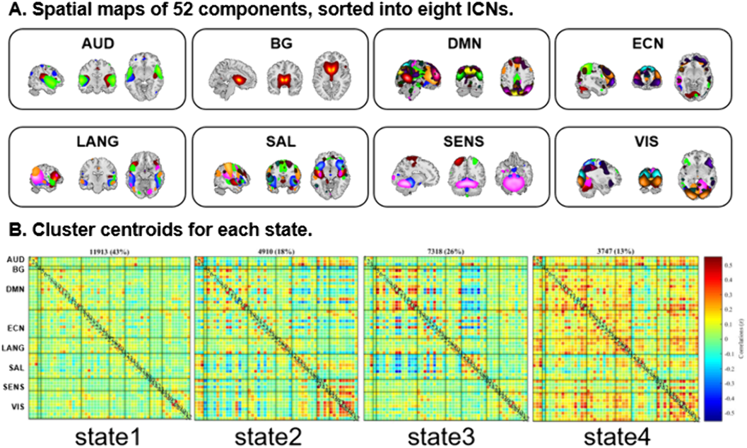
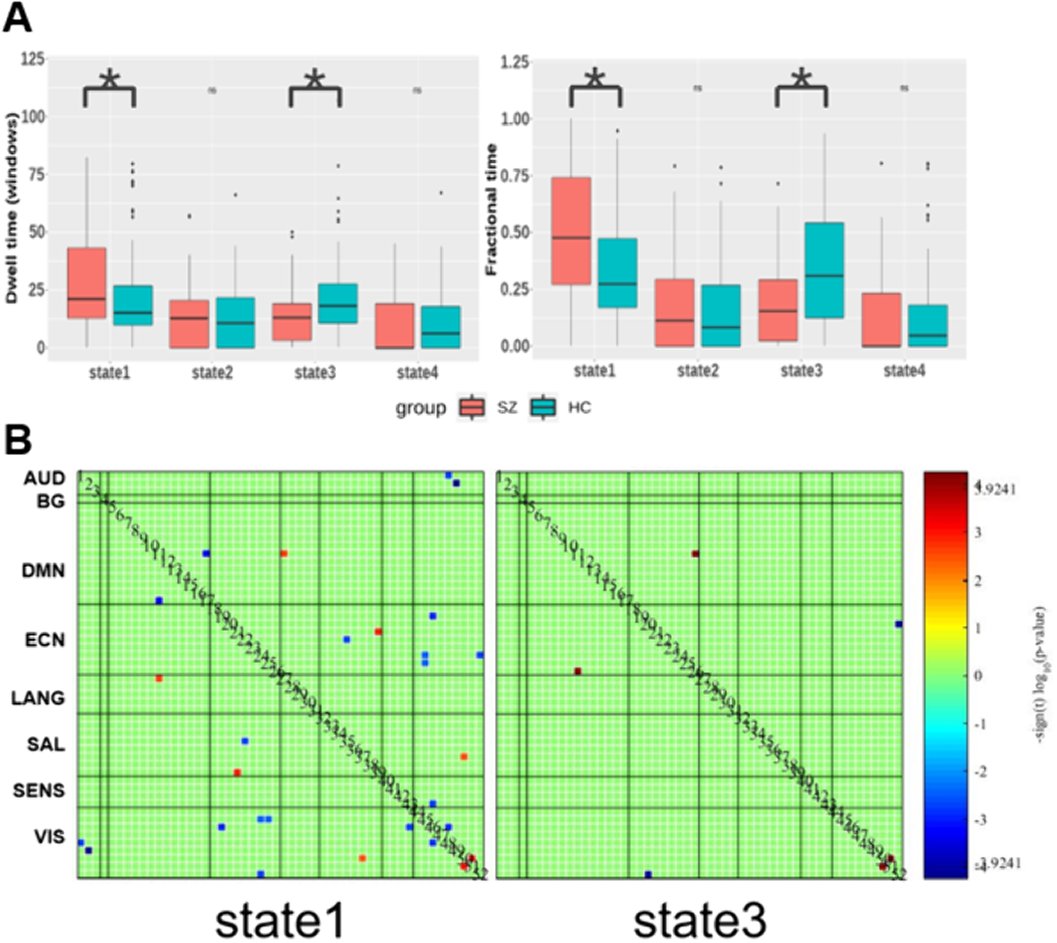
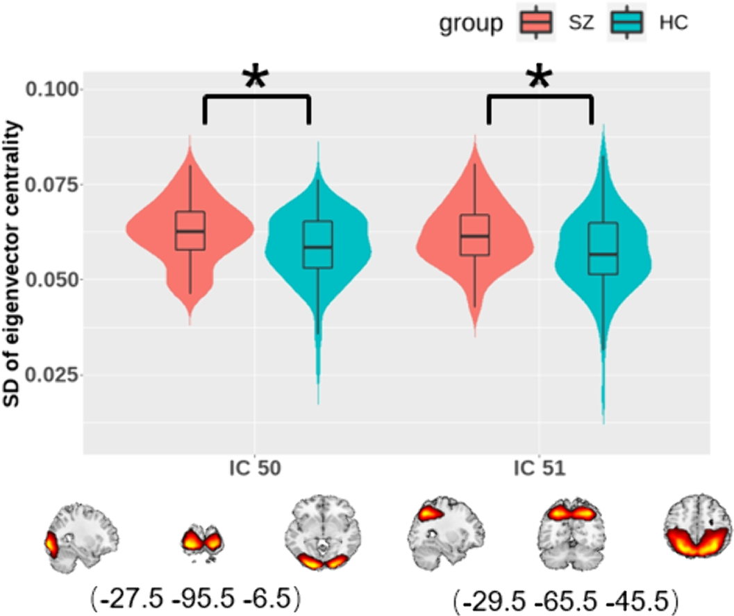
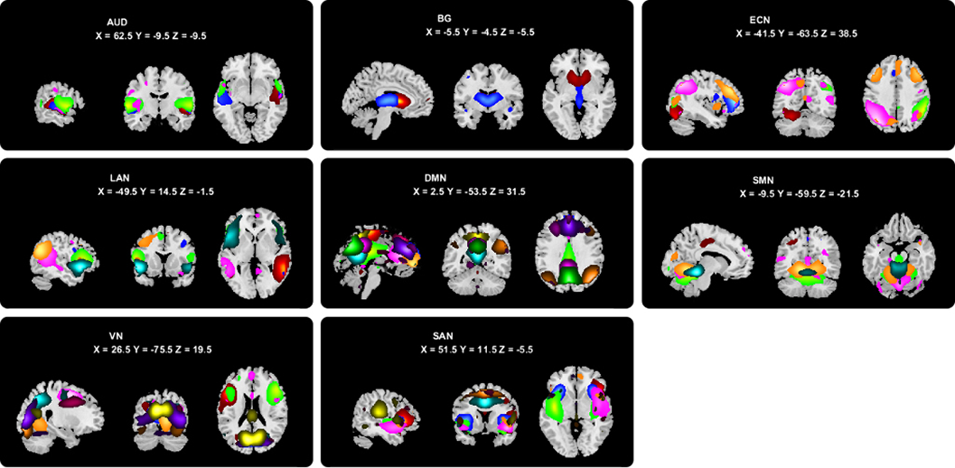
Methods
Using magnetic resonance imaging for 83 SZ patients and 83 matched healthy controls and independent component analysis, 52 independent components (ICs) were identified as nodes and assigned into eight intrinsic connectivity networks (Figure 1A). Subsequently, we established dFC matrices and clustered them into four discrete states (Figure 1B) and three state transition metrics were obtained. To further explore the changes in the centrality of each component, eigenvector centrality (EC) was calculated and its time-varying was evaluated.
Results
Compared to controls with FDR correction, we found that patients had more mean dwell times and fractional time in state 1 (P=0.0081 and P=0.0018), mainly with hypoconnectivity between auditory and visual network and other networks and hyperconnectivity between language and default-mode network (DMN). While, patients had less dwell times and fractional time in state 3 (P=0.0018 and P=0.0009), and decreased FC between visual network and executive control network (ECN) and increased FC between ECN and DMN than controls (Figure 2).
EC statistics showed that SZs displayed increased temporal dynamics in visual-related regions (Figure 3).
Conclusions
SZ was mainly manifested as altered dFC and temporal variability of nodal centrality in auditory and visual networks.
Though schizophrenia (SZ) and obsessive-compulsive disorder (OCD) are conceptualized as distinct clinical entities, they do have notable symptom overlap and a tight association. Graph-theoretical analysis of the brain connectome provides more indicators to describe the functional organization of the brain, which may help us understand the shared and disorder-specific neural basis of the two disorders.
Objectives
To explore the static and dynamic topological organization of OCD and SZ as well as the relationship between topological metrics and clinical variables.
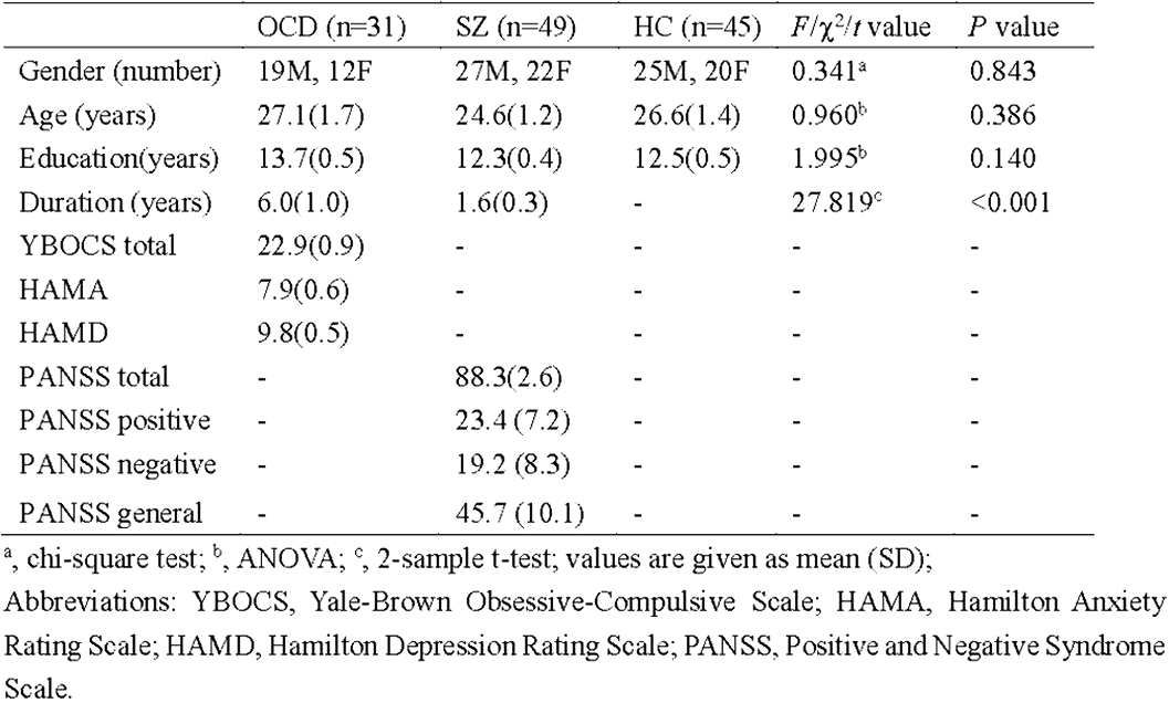
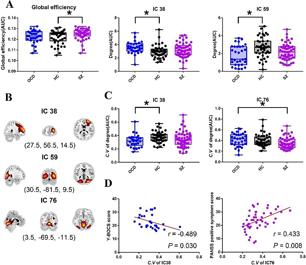
Methods
Resting state functional magnetic resonance imaging data of 31 OCD patients, 49 SZ patients, and 45 healthy controls (HC) were involved in this study (Table 1). Using independent component analysis to obtain independent components (ICs) (Figure 1), which were defined as nodes for static and dynamic topological analysis.
Results
Static analysis showed the global efficiency of SZ was higher than HC. For nodal degree centrality, OCD exhibited decreased degree centrality in IC59 (located in visiual network) (P = 0.03) and increased degree centrality in IC38 (located in salience network) (P = 0.002) compared with HC. Dynamic analysis showed OCD exhibited decreased dynamics of degree centrality in IC38 (P = 0.003) compared with HC, which showed a negative correlation with clinical scores in OCD. While SZ showed decreased dynamics of degree centrality in IC76 (located in sensory motor network) compared with OCD (P=0.009), which showed a positive correlation with clinical scores in SZ (Figure 2).
Conclusions
These changes are suggestive of disorder-specific alternation of static and dynamic brain topological organization in OCD and SZ.
Obsessive-compulsive disorder (OCD) and schizophrenia (SZ) are both severe psychiatric disorders. Though these two disorders have distinct typical symptoms, there are partial polygenic overlap and comorbidity between the two disorders. However, few studies have explored the shared and disorder-specific brain function underlying the neural pathophysiology of the two disorders, especially in the aspect of dynamics.
Objectives
To explore the abnormal characteristics of the dynamic functional connectivity (dFC) in OCD and SZ as well as the association between dFC metrics and symptom severity.
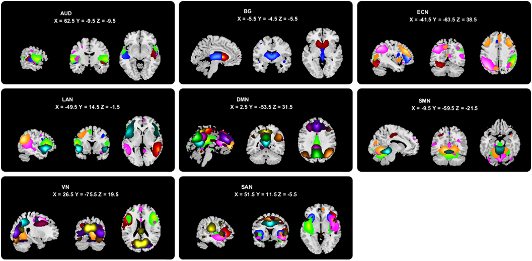
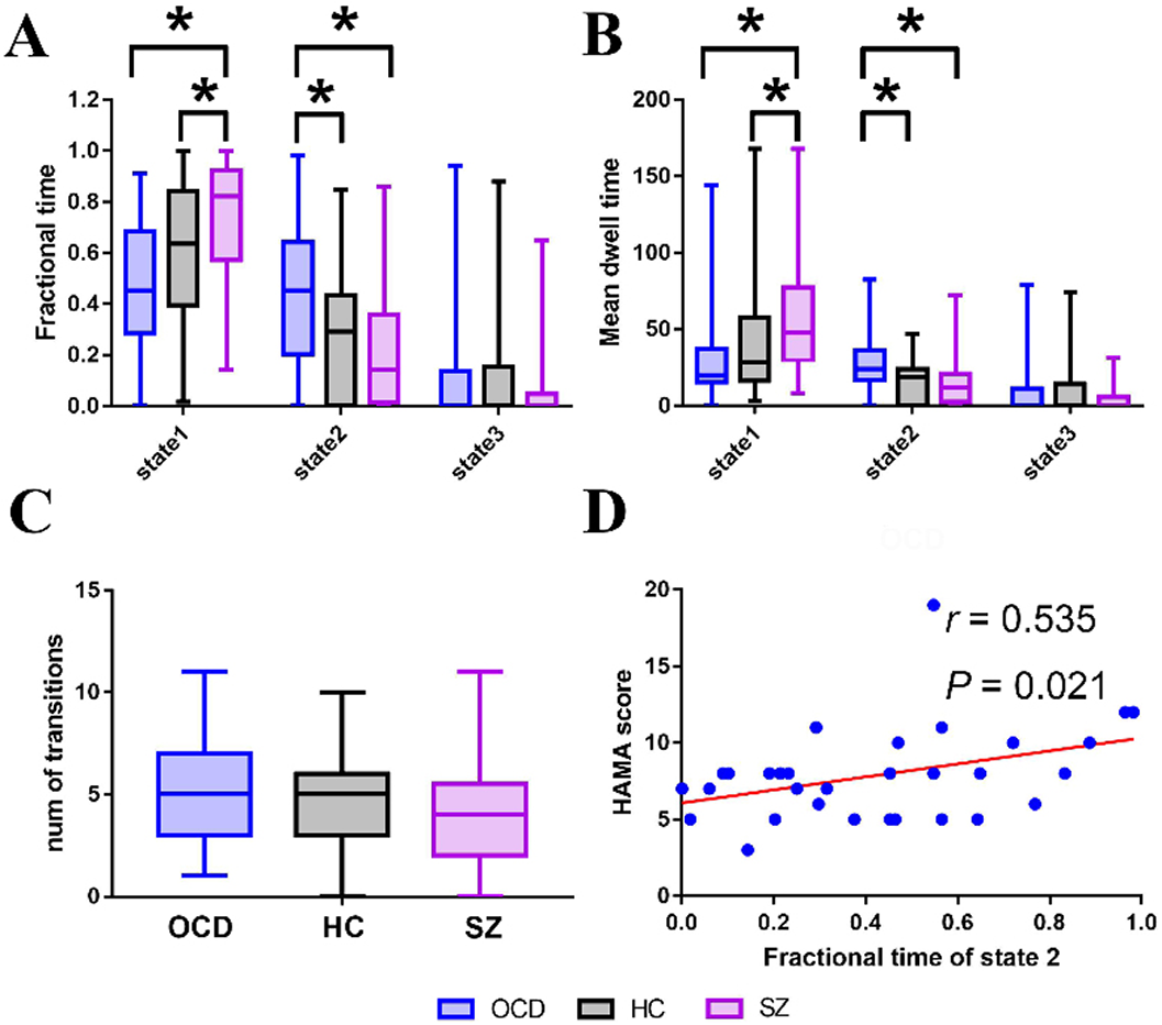
Methods
The resting state functional magnetic resonance imaging data of 31 patients with OCD, 49 patients with SZ, and 45 healthy controls were analyzed using independent component analysis to obtain independent components (ICs) and assigned them into eight brain networks (Figure 1), then used the sliding-window approach to generate dFC matrices. Using k-means clustering, we obtained three reoccurring dFC states (Figure 2), and state transition metrics were obtained
Results
In a sparsely connected state (state 1), SZ showed both increased fractional time and mean dwell time than controls (P=0.047 and P=0.033) and OCD (P=0.001 and P=0.003). In a state characterized by negative FC between networks (state 2), OCD showed both increased fractional time and mean dwell time than controls (P=0.032 and P=0.013) and SZ (P=0.005 and P=0.003). Moreover, the fractional time of state 2 was positively correlated with anxiety scores in OCD (r=0.535, P=0.021, FDR corrected) (Figure 3).
Conclusions
OCD and SZ patients showed distinct alternations of brain functional dynamics.
Schizotypy refers to schizophrenia-like traits below the clinical threshold in the general population. The pathological development of schizophrenia has been postulated to evolve from the initial coexistence of ‘brain disconnection’ and ‘brain connectivity compensation’ to ‘brain connectivity decompensation’.
Methods
In this study, we examined the brain connectivity changes associated with schizotypy by combining brain white matter structural connectivity, static and dynamic functional connectivity analysis of diffusion tensor imaging data and resting-state functional magnetic resonance imaging data. A total of 87 participants with a high level of schizotypal traits and 122 control participants completed the experiment. Group differences in whole-brain white matter structural connectivity probability, static mean functional connectivity strength, dynamic functional connectivity variability and stability among 264 brain sub-regions of interests were investigated.
Results
We found that individuals with high schizotypy exhibited increased structural connectivity probability within the task control network and within the default mode network; increased variability and decreased stability of functional connectivity within the default mode network and between the auditory network and the subcortical network; and decreased static mean functional connectivity strength mainly associated with the sensorimotor network, the default mode network and the task control network.
Conclusions
These findings highlight the specific changes in brain connectivity associated with schizotypy and indicate that both decompensatory and compensatory changes in structural connectivity within the default mode network and the task control network in the context of whole-brain functional disconnection may be an important neurobiological correlate in individuals with high schizotypy.
Accumulating studies have found structural and functional abnormalities of the striatum in bipolar disorder (BD) and major depressive disorder (MDD). However, changes in intrinsic brain functional connectivity dynamics of striato-cortical circuitry have not been investigated in BD and MDD. This study aimed to investigate the shared and specific patterns of dynamic functional connectivity (dFC) variability of striato-cortical circuitry in BD and MDD.
Methods
Brain resting-state functional magnetic resonance imaging data were acquired from 128 patients with unmedicated BD II (current episode depressed), 140 patients with unmedicated MDD, and 132 healthy controls (HCs). Six pairs of striatum seed regions were selected: the ventral striatum inferior (VSi) and the ventral striatum superior (VSs), the dorsal-caudal putamen (DCP), the dorsal-rostral putamen (DRP), and the dorsal caudate and the ventral-rostral putamen (VRP). The sliding-window analysis was used to evaluate dFC for each seed.
Results
Both BD II and MDD exhibited increased dFC variability between the left DRP and the left supplementary motor area, and between the right VRP and the right inferior parietal lobule. The BD II had specific increased dFC variability between the right DCP and the left precentral gyrus compared with MDD and HCs. The MDD had increased dFC variability between the left VSi and the left medial prefrontal cortex compared with BD II and HCs.
Conclusions
The patients with BD and MDD shared common dFC alteration in the dorsal striatal-sensorimotor and ventral striatal-cognitive circuitries. The patients with MDD had specific dFC alteration in the ventral striatal-affective circuitry.
Recommend this
Email your librarian or administrator to recommend adding this to your organisation's collection.